
 Actinin-α3 (E25) polyclonal antibody
  Datasheet
 COA
COA MSDS
MSDS SHIP
SHIP
Product Name :
Actinin-α3 (E25) polyclonal antibody Background :
The spectrin gene family encodes a diverse group of cytoskeletal proteins that include spectrins, dystrophins and α-actinins. There are four tissue-specific α-actinins, namely α-actinin-1, α-actinin-2, α-actinin-3 and α-actinin-4, which are localized to muscle and non-muscle cells, including skeletal, cardiac and smooth muscle cells, as well as within the cytoskeleton. Each α-actinin protein contains one Actin-binding domain, two calponin-homology domains, two EF-hand domains and four spectrin repeats, through which they function as bundling proteins that can cross-link F-Actin, thus anchoring Actin to a variety of intracellular structures. Defects in the gene encoding α-actinin-4 are the cause of focal segmental glomerulosclerosis 1 (FSGS1), a common renal lesion characterized by decreasing kidney function and, ultimately, renal failure. Product :
Rabbit IgG, 1mg/ml in PBS with 0.02% sodium azide, 50% glycerol, pH7.2 Storage&Stability :
Store at 4°C short term. Aliquot and store at -20°C long term. Avoid freeze-thaw cycles. Specificity :
Actinin-α3 (E25) polyclonal antibody detects endogenous levels of actinin-α3 protein, this antibody does not cross-react with other α-actinin isoforms. Immunogen :
Synthetic peptide, corresponding to the N-terminus of Human Actinin-α3. Conjugate :
Unconjugated Modification :
Unmodification
-
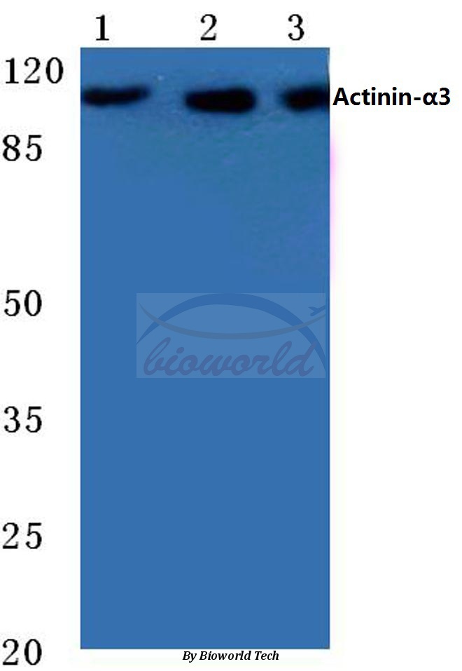 Western blot (WB) analysis of Actinin-α3 (E25) polyclonal antibody at 1:500 dilution Lane1:Hela cell lysate Lane2:Mouse muscle tissue lysate Lane3:Rat muscel tissue lysate
 -
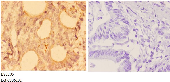 Immunohistochemistry (IHC) analyzes of Actinin-α3 (E25) pAb in paraffin-embedded human colon carcinoma tissue at 1:50,showing cytoplasmic and nucleus staining.Negative control (the right)Using PBS instead of primary antibody, secondary antibody is Goat Anti-Rabbit IgG-biotin followed by avidin-peroxidase.
 -
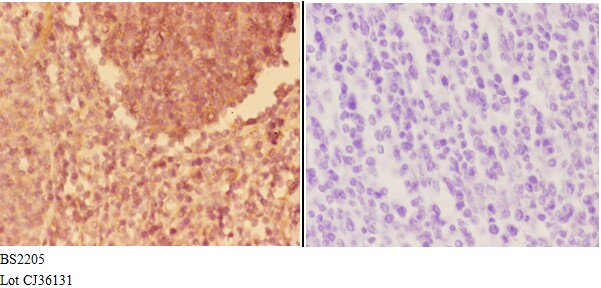 Immunohistochemistry (IHC) analyzes of Actinin-α3 (E25) pAb in paraffin-embedded human colon carcinoma tissue at 1:50,showing cytoplasmic and nucleus staining.Negative control (the right)Using PBS instead of primary antibody, secondary antibody is Goat Anti-Rabbit IgG-biotin followed by avidin-peroxidase.
Bioworld Biotech only provide peptides for our antibodies and do not provide additional peptide customization services.
Price/Size :
USD 368/1mg/vial

Tips:
For phospho antibody, we provide phospho peptide(0.5mg) and non-phospho peptide(0.5mg).Describe :
Blocking peptides are peptides that bind specifically to the target antibody and block antibody binding. These peptide usually contains the epitope recognized by the antibody. Antibodies bound to the blocking peptide no longer bind to the epitope on the target protein. This mechanism is useful when non-specific binding is an issue, for example, in Western blotting (WB) and Immunohistochemistry (IHC). By comparing the staining from the blocked antibody versus the antibody alone, one can see which staining is specific; Specific binding will be absent from the western blot or IHC performed with the neutralized antibody.Formula:
Synthetic peptide was lyophilized with 100% acetonitrile and is supplied as a powder. Reconstitute with 0.1 ml DI water for a final concentration of 10 mg/ml.The purity is >90%,tested by HPLC and MS.
Storage:
The freeze-dried powder is more stable. For short time at 2-8°C. For long term storage store at -20°C.
Note :
This product is for research use only (RUO only). Not for use in diagnostic or therapeutic procedures.