
 PKC α (phospho-T638) polyclonal antibody
  Datasheet
 COA
COA MSDS
MSDS SHIP
SHIP
Product Name :
PKC α (phospho-T638) polyclonal antibody Background :
Members of the protein kinase C (PKC) family play a key regulatory role in a variety of cellular functions including cell growth and differentiation, gene expression, hormone secretion and membrane function. PKCs were originally identified as serine/threonine protein kinases whose activity was dependent on calcium and phospholipids. Diacylglycerols (DAG) and tumor promoting phorbol esters bind to and activate PKC. PKCs can be subdivided into at least two major classes including conventional (c) PKC isoforms (α, βI, βII and γ) and novel (n) PKC isoforms (δ, ε, ζ, η and θ). Patterns of expression for each PKC isoform differ among tissues and PKC family members exhibit clear differences in their cofactor dependencies. For instance, the kinase activities of nPKC δ and ε are independent of Ca2+. Product :
Rabbit IgG, 1mg/ml in PBS with 0.02% sodium azide, 50% glycerol, pH7.2 Storage&Stability :
Store at 4°C short term. Aliquot and store at -20°C long term. Avoid freeze-thaw cycles. Specificity :
PKC α (phospho-T638) polyclonal antibody detects endogenous levels of PKC α protein when phosphorylated at Thr638. Immunogen :
Synthetic phosphopeptide derived from human PKC α around the phosphorylation site of Threonine 638. Conjugate :
Unconjugated Modification :
Phosphorylation
-
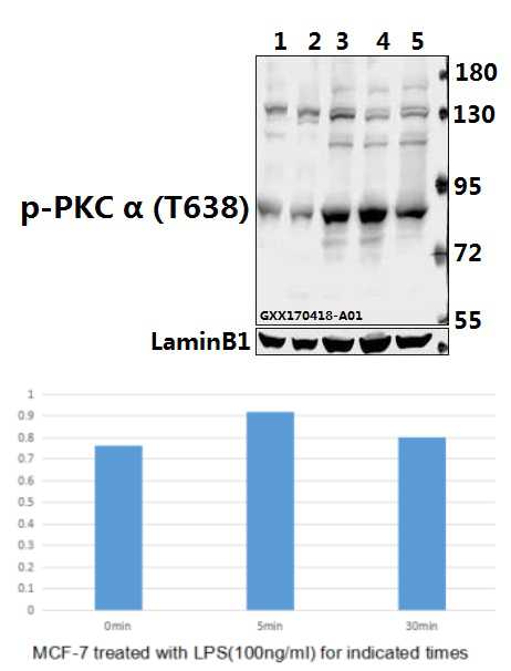 Western blot (WB) analysis of PKC α (phospho-T638) polyclonal antibody at 1:500 dilution Lane1:PC12 whole cell lysate(40ug) Lane2:CT26 whole cell lysate(40ug) Lane3:MCF-7 treated with LPS(100ng/ml) for 30 minutes whole cell lysate Lane4:MCF-7 treated with LPS(100ng/ml) for 5 minutes whole cell lysate Lane5:MCF-7 whole cell lysate
 -
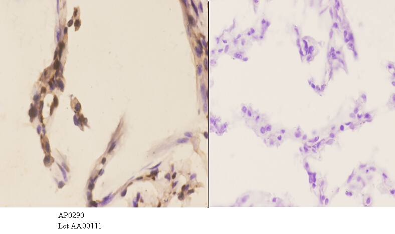 Immunohistochemistry (IHC) analyzes of p-PKC α (T638) pAb in paraffin-embedded human lung carcinoma tissue at 1:50.showing cytoplasmic and nucleus staining. Negative control (the right)Using PBS instead of primary antibody, secondary antibody is Goat Anti-Rabbit IgG-biotin followed by avidin-peroxidase.
 -
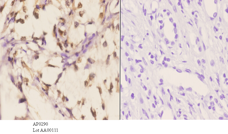 Immunohistochemistry (IHC) analyzes of p-PKC α (T638) pAb in paraffin-embedded human lung carcinoma tissue at 1:50.showing cytoplasmic and nucleus staining. Negative control (the right)Using PBS instead of primary antibody, secondary antibody is Goat Anti-Rabbit IgG-biotin followed by avidin-peroxidase.
Norisoboldine inhibits the production of interleukin-6 in fibroblast-like synoviocytes from adjuvant arthritis rats through PKC/MAPK/NF-κB-p65/CREB pathways
PMCID: Pubmed No.:22473817
Reduced expression of annexin A1 promotes gemcitabine and 5-fluorouracil drug resistance of human pancreatic cancer
PMCID: Pubmed No.:31124054
Bioworld Biotech only provide peptides for our antibodies and do not provide additional peptide customization services.
Price/Size :
USD 368/1mg/vial

Tips:
For phospho antibody, we provide phospho peptide(0.5mg) and non-phospho peptide(0.5mg).Describe :
Blocking peptides are peptides that bind specifically to the target antibody and block antibody binding. These peptide usually contains the epitope recognized by the antibody. Antibodies bound to the blocking peptide no longer bind to the epitope on the target protein. This mechanism is useful when non-specific binding is an issue, for example, in Western blotting (WB) and Immunohistochemistry (IHC). By comparing the staining from the blocked antibody versus the antibody alone, one can see which staining is specific; Specific binding will be absent from the western blot or IHC performed with the neutralized antibody.Formula:
Synthetic peptide was lyophilized with 100% acetonitrile and is supplied as a powder. Reconstitute with 0.1 ml DI water for a final concentration of 10 mg/ml.The purity is >90%,tested by HPLC and MS.
Storage:
The freeze-dried powder is more stable. For short time at 2-8°C. For long term storage store at -20°C.
Note :
This product is for research use only (RUO only). Not for use in diagnostic or therapeutic procedures.