
 AKT (phospho-T450) polyclonal antibody
  Datasheet
 COA
COA MSDS
MSDS SHIP
SHIP
Product Name :
AKT (phospho-T450) polyclonal antibody Background :
AKT, also known as protein kinase B (PKB), is a 57 kDa serine/threonine protein kinase. There are three mammalian isoforms of Akt: AKT1 (PKB alpha), AKT2 (PKB beta) and AKT3 (PKB gamma) with AKT2 and AKT3 being approximately 82% identical with the AKT1 isoform. Each isoform has a pleckstrin homology (PH) domain, a kinase domain and a carboxy terminal regulatory domain. AKT was originally cloned from the retrovirus AKT8, and is a key regulator of many signal transduction pathways. Its tight control over cell proliferation and cell viability are manifold; overexpression or inappropriate activation of AKT has been seen in many types of cancer. Product :
Rabbit IgG, 1mg/ml in PBS with 0.02% sodium azide, 50% glycerol, pH7.2 Storage&Stability :
Store at 4°C short term. Aliquot and store at -20°C long term. Avoid freeze-thaw cycles. Specificity :
p-AKT (T450) polyclonal antibody detects endogenous levels of AKT1 only when phosphorylated at Thr450. This antibody also recognizes AKT2 and AKT3 when phosphorylated at the corresponding residues. Immunogen :
Synthetic phosphopeptide derived from human AKT1 around the phosphorylation site of Threonine 450. Conjugate :
Unconjugated Modification :
Phosphorylated
-
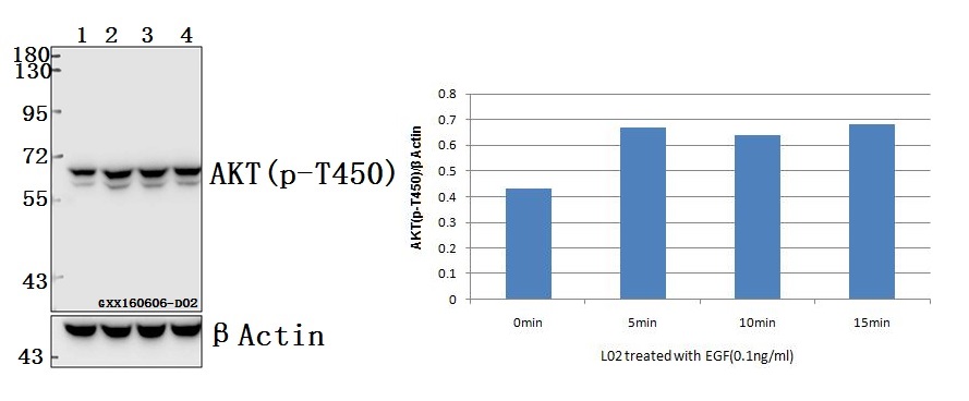 Western blot (WB) analysis of AKT (phospho-T450) polyclonal antibody at 1:500 dilution Lane1:L02 whole cell lysate Lane2:L02 treated with EGF(0.1ng/ml,5min) whole cell lysate Lane3:L02 treated with EGF(0.1ng/ml,10min) whole cell lysate Lane4:L02 treated with EGF(0.1ng/ml,15min) whole cell lysate
 -
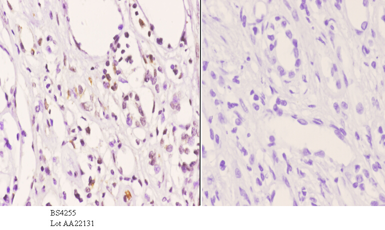 Immunohistochemistry (IHC) analyzes of p-AKT (T450) pAb in paraffin-embedded human kidney carcinoma tissue at 1:50.showing cytoplasmic and nucleus staining. Negative control (the right)Using PBS instead of primary antibody, secondary antibody is Goat Anti-Rabbit IgG-biotin followed by avidin-peroxidase.
MiR-205 inhibits cell apoptosis by targeting phosphatase and tensin homolog deleted on chromosome ten in endometrial cancer ishikawa cells
PMCID: Pubmed No.:24929707
Nicorandil inhibits oxidative stress and amyloid-β precursor protein processing in SH-SY5Y cells overexpressing APPsw.
PMCID: Pubmed No.:25932125
Bioworld Biotech only provide peptides for our antibodies and do not provide additional peptide customization services.
Price/Size :
USD 368/1mg/vial

Tips:
For phospho antibody, we provide phospho peptide(0.5mg) and non-phospho peptide(0.5mg).Describe :
Blocking peptides are peptides that bind specifically to the target antibody and block antibody binding. These peptide usually contains the epitope recognized by the antibody. Antibodies bound to the blocking peptide no longer bind to the epitope on the target protein. This mechanism is useful when non-specific binding is an issue, for example, in Western blotting (WB) and Immunohistochemistry (IHC). By comparing the staining from the blocked antibody versus the antibody alone, one can see which staining is specific; Specific binding will be absent from the western blot or IHC performed with the neutralized antibody.Formula:
Synthetic peptide was lyophilized with 100% acetonitrile and is supplied as a powder. Reconstitute with 0.1 ml DI water for a final concentration of 10 mg/ml.The purity is >90%,tested by HPLC and MS.
Storage:
The freeze-dried powder is more stable. For short time at 2-8°C. For long term storage store at -20°C.
Note :
This product is for research use only (RUO only). Not for use in diagnostic or therapeutic procedures.