
 p53 (Ab-20) polyclonal antibody
  Datasheet
 COA
COA MSDS
MSDS SHIP
SHIP
Product Name :
p53 (Ab-20) polyclonal antibody Background :
The p53 tumor suppressor protein plays a major role in cellular response to DNA damage and other genomic aberrations. Activation of p53 can lead to either cell cycle arrest and DNA repair or apoptosis. p53 is phosphorylated at multiple sites in vivo and by several different protein kinases in vitro. DNA damage induces phosphorylation of p53 at Ser15 and Ser20 and leads to a reduced interaction between p53 and its negative regulator, the oncoprotein MDM2. MDM2 inhibits p53 accumulation by targeting it for ubiquitination and proteasomal degradation. p53 can be phosphorylated by ATM, ATR, and DNA-PK at Ser15 and Ser37. Phosphorylation impairs the ability of MDM2 to bind p53, promoting both the accumulation and activation of p53 in response to DNA damage. Chk2 and Chk1 can phosphorylate p53 at Ser20, enhancing its tetramerization, stability, and activity. p53 is phosphorylated at Ser392 in vivo and by CAK in vitro. Phosphorylation of p53 at Ser392 is increased in human tumors and has been reported to influence the growth suppressor function, DNA binding, and transcriptional activation of p53. p53 is phosphorylated at Ser6 and Ser9 by CK1δ and CK1ε both in vitro and in vivo. Phosphorylation of p53 at Ser46 regulates the ability of p53 to induce apoptosis. Acetylation of p53 is mediated by p300 and CBP acetyltransferases. Inhibition of deacetylation suppressing MDM2 from recruiting HDAC1 complex by p19 (ARF) stabilizes p53. Acetylation appears to play a positive role in the accumulation of p53 protein in stress response. Following DNA damage, human p53 becomes acetylated at Lys382 (Lys379 in mouse) in vivo to enhance p53-DNA binding. Deacetylation of p53 occurs through interaction with the SIRT1 protein, a deacetylase that may be involved in cellular aging and the DNA damage response. Product :
Rabbit IgG, 1mg/ml in PBS with 0.02% sodium azide, 50% glycerol, pH7.2. Storage&Stability :
Store at 4°C short term. Aliquot and store at -20°C long term. Avoid freeze-thaw cycles. Specificity :
p53 (Ab-20) polyclonal antibody detects endogenous levels of p53 protein. Immunogen :
Synthetic peptide, corresponding to Human p53. Conjugate :
Unconjugated Modification :
Unmodified
-
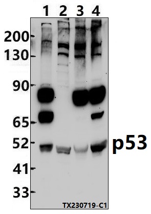 Western blot (WB) analysis of p53 (Ab-20) polyclonal antibody at 1:500 dilution Lane1:HEK293T whole cell lysate(30ug) Lane2:3T3-L1 whole cell lysate(30ug) Lane3:EC9706 whole cell lysate(30ug) Lane4:Pancl whole cell lysate(30ug)
 -
A critical role for interferon regulatory factor 9 in cerebral ischemic stroke.
PMCID: Pubmed No.:25186738
MCM2 is a therapeutic target of lovastatin in human non-small cell lung carcinomas
PMCID: Pubmed No.:25738322
A critical role for interferon regulatory factor 9 in cerebral ischemic stroke.
PMCID: Pubmed No.:25186738
Bioworld Biotech only provide peptides for our antibodies and do not provide additional peptide customization services.
Price/Size :
USD 368/1mg/vial

Tips:
For phospho antibody, we provide phospho peptide(0.5mg) and non-phospho peptide(0.5mg).Describe :
Blocking peptides are peptides that bind specifically to the target antibody and block antibody binding. These peptide usually contains the epitope recognized by the antibody. Antibodies bound to the blocking peptide no longer bind to the epitope on the target protein. This mechanism is useful when non-specific binding is an issue, for example, in Western blotting (WB) and Immunohistochemistry (IHC). By comparing the staining from the blocked antibody versus the antibody alone, one can see which staining is specific; Specific binding will be absent from the western blot or IHC performed with the neutralized antibody.Formula:
Synthetic peptide was lyophilized with 100% acetonitrile and is supplied as a powder. Reconstitute with 0.1 ml DI water for a final concentration of 10 mg/ml.The purity is >90%,tested by HPLC and MS.
Storage:
The freeze-dried powder is more stable. For short time at 2-8°C. For long term storage store at -20°C.
Note :
This product is for research use only (RUO only). Not for use in diagnostic or therapeutic procedures.