
 Glut1 polyclonal antibody
  Datasheet
 COA
COA MSDS
MSDS SHIP
SHIP
Product Name :
Glut1 polyclonal antibody Background :
Glucose is fundamental to the metabolism of mammalian cells. Its passage across cell membranes is mediated by a family of transporters termed glucose transporters or Gluts. In adipose and muscle tissue, insulin stimulates a rapid and dramatic increase in glucose uptake, which is largely due to the redistribution of the insulin-inducible glucose transporter, Glut4. In response to insulin, Glut4 is quickly shuttled from an intracellular storage site to the plasma membrane, where it binds glucose. In contrast, the ubiquitously expressed glucose transporter Glut1 is constitutively targeted to the plasma membrane, and shows a much less dramatic translocation in response to insulin. Glut1 and Glut4 are twelve-pass transmembrane proteins (12TM) whose carboxy-termini may dictate their cellular localization. Aberrant Glut4 expression has been suggested to contribute to such maladies as obesity and diabetes. Glut4 null mice have shown that while functional Glut4 protein is not required for maintaining normal glucose levels, it is necessary for sustained growth, normal cellular glucose, fat metabolism and prolonged longevity. Product :
Rabbit IgG, 1mg/ml in PBS with 0.02% sodium azide, 50% glycerol, pH7.2 Storage&Stability :
Store at +4°C after thawing. Aliquot store at -20°C or -80°C. Avoid repeated freeze / thaw cycles. Specificity :
Glut1 polyclonal antibody detects endogenous levels of Glut1 protein. Immunogen :
Peptide. Conjugate :
Unconjugated Modification :
Unmodification
-
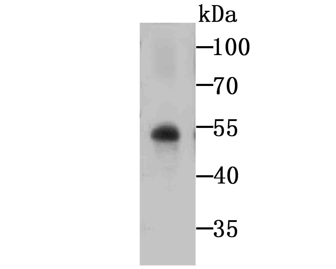 Western blot analysis of Glut1 on human placenta tissue lysates using anti-Glut1 antibody at 1/1,000 dilution.
 -
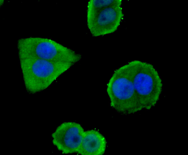 ICC staining Glut1 in Hela cells (green). The nuclear counter stain is DAPI (blue). Cells were fixed in paraformaldehyde, permeabilised with 0.25% Triton X100/PBS.
Pyruvate kinase M2 accelerates pro-inflammatory cytokine secretion and cell proliferation induced by lipopolysaccharide in colorectal cancer
PMCID: Pubmed No.:25778902
GRP78 is implicated in the modulation of tumor aerobic glycolysis by promoting autophagic degradation of IKKβ
PMCID: Pubmed No.:25748049
Oncogenic TRIB2 interacts with and regulates PKM2 to promote aerobic glycolysis and lung cancer cell procession
PMCID: Pubmed No.:35790734
Bioworld Biotech only provide peptides for our antibodies and do not provide additional peptide customization services.
Price/Size :
USD 368/1mg/vial

Tips:
For phospho antibody, we provide phospho peptide(0.5mg) and non-phospho peptide(0.5mg).Describe :
Blocking peptides are peptides that bind specifically to the target antibody and block antibody binding. These peptide usually contains the epitope recognized by the antibody. Antibodies bound to the blocking peptide no longer bind to the epitope on the target protein. This mechanism is useful when non-specific binding is an issue, for example, in Western blotting (WB) and Immunohistochemistry (IHC). By comparing the staining from the blocked antibody versus the antibody alone, one can see which staining is specific; Specific binding will be absent from the western blot or IHC performed with the neutralized antibody.Formula:
Synthetic peptide was lyophilized with 100% acetonitrile and is supplied as a powder. Reconstitute with 0.1 ml DI water for a final concentration of 10 mg/ml.The purity is >90%,tested by HPLC and MS.
Storage:
The freeze-dried powder is more stable. For short time at 2-8°C. For long term storage store at -20°C.
Note :
This product is for research use only (RUO only). Not for use in diagnostic or therapeutic procedures.