
 GATA4 polyclonal antibody
  Datasheet
 COA
COA MSDS
MSDS SHIP
SHIP
Product Name :
GATA4 polyclonal antibody Background :
Members of the GATA family share a conserved zinc finger DNA-binding domain and are capable of binding the WGATAR consensus sequence. GATA-1 is erythroid-specific and is responsible for the regulated transcription of erythroid genes. It is an essential component in the generation of the erythroid lineage. GATA-2 is expressed in embryonic brain and liver, HeLa and endothelial cells, as well as in erythroid cells. Studies with a modified GATA consensus sequence, AGATCTTA, have shown that GATA-2 and GATA-3 recognize this mutated consensus while GATA-1 has poor recognition of this sequence. This indicates broader regulatory capabilities of GATA-2 and GATA-3 than GATA-1. GATA-3 is highly expressed in T lymphocytes. GATA-4, GATA-5 and GATA-6 comprise a subfamily of transcription factors. Both GATA-4 and GATA-6 are found in heart, pancreas and ovary; lung and liver tissues exhibit GATA-6, but not GATA-4 expression. GATA-5 expression has been observed in differentiated heart and gut tissues and is present throughout the course of development in the heart. Although expression patterns of the various GATA transcription factors may overlap, it is not yet apparent how the GATA factors are able to discriminate in binding their appropriate target sites. Product :
Rabbit IgG, 1mg/ml in PBS with 0.02% sodium azide, 50% glycerol, pH7.2 Storage&Stability :
Store at +4°C after thawing. Aliquot store at -20°C or -80°C. Avoid repeated freeze / thaw cycles. Specificity :
GATA4 polyclonal antibody detects endogenous levels of GATA4 protein. Immunogen :
recombinant protein Conjugate :
Unconjugated Modification :
Unmodification
-
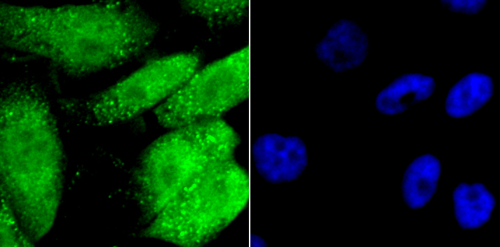 ICC staining GATA4 in PC-3M cells (Green). The nuclear counter stain is DAPI (blue). Cells were fixed in paraformaldehyde, permeabilised with 0.25% Triton X100/PBS.
 -
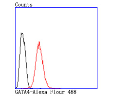 Flow cytometric analysis of Hela cells with GATA4 antibody at 1/50 dilution (red) compared with an unlabelled control (cells without incubation with primary antibody; black). Alexa Fluor 488-conjugated goat anti rabbit IgG was used as the secondary antibody.
Bioworld Biotech only provide peptides for our antibodies and do not provide additional peptide customization services.
Price/Size :
USD 368/1mg/vial

Tips:
For phospho antibody, we provide phospho peptide(0.5mg) and non-phospho peptide(0.5mg).Describe :
Blocking peptides are peptides that bind specifically to the target antibody and block antibody binding. These peptide usually contains the epitope recognized by the antibody. Antibodies bound to the blocking peptide no longer bind to the epitope on the target protein. This mechanism is useful when non-specific binding is an issue, for example, in Western blotting (WB) and Immunohistochemistry (IHC). By comparing the staining from the blocked antibody versus the antibody alone, one can see which staining is specific; Specific binding will be absent from the western blot or IHC performed with the neutralized antibody.Formula:
Synthetic peptide was lyophilized with 100% acetonitrile and is supplied as a powder. Reconstitute with 0.1 ml DI water for a final concentration of 10 mg/ml.The purity is >90%,tested by HPLC and MS.
Storage:
The freeze-dried powder is more stable. For short time at 2-8°C. For long term storage store at -20°C.
Note :
This product is for research use only (RUO only). Not for use in diagnostic or therapeutic procedures.