
 Galectin 3 polyclonal antibody
  Datasheet
 COA
COA MSDS
MSDS SHIP
SHIP
Product Name :
Galectin 3 polyclonal antibody Background :
Galectins are a family of soluble b-galactoside-binding animal lectins that modulate cell-to-cell adhesion and cell-to-extracellular matrix (ECM) inter- actions and play a role in tumor progression, pre-mRNA splicing and apop-tosis. The galectin-3 protein, also known as Mac-2, hMac-2, GALBP, CBP35 or LGALS3, contains a single carbohydrate binding domain, which binds galactose-containing glycoconjugates. Galectin-3 is expressed in colonic and intestinal epithelium, inflammatory macrophages, papillary and follicular carcinomas, neoplastic astrocytes and some B and T lymphocytes. Upregulated expression of galectin-3 is involved in cancer progression and metastasis. Galectin-3 mediates the endocytosis of β1 Integrins in a lactose-dependent manner and is associated with thyroid malignancy and Crohn's disease. It may also be used as a marker for diagnosing cases involving Hurthle cell adenomas and carcinomas. Product :
Rabbit IgG, 1mg/ml in PBS with 0.02% sodium azide, 50% glycerol, pH7.2 Storage&Stability :
Store at +4℃ after thawing. Aliquot store at -20℃. Avoid repeated freeze / thaw cycles. Specificity :
Galectin 3 polyclonal antibody detects endogenous levels of Galectin 3 protein. Immunogen :
Recombinant protein within Human Galectin 3 aa 1-180. Conjugate :
Unconjugated Modification :
Unmodification
-
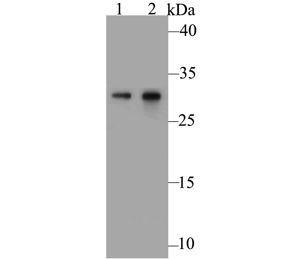 Western blot analysis of Galectin 3 on different lysates. Proteins were transferred to a PVDF membrane and blocked with 5% BSA in PBS for 1 hour at room temperature. The primary antibody was used at a 1:500 dilution in 5% BSA at room temperature for 2 hours. Goat Anti-Rabbit IgG - HRP Secondary Antibody (HA1001) at 1:5,000 dilution was used for 1 hour at room temperature.Positive control: Lane 1: SiHa cell lysateLane 2: Mouse colon tissue lysate
 -
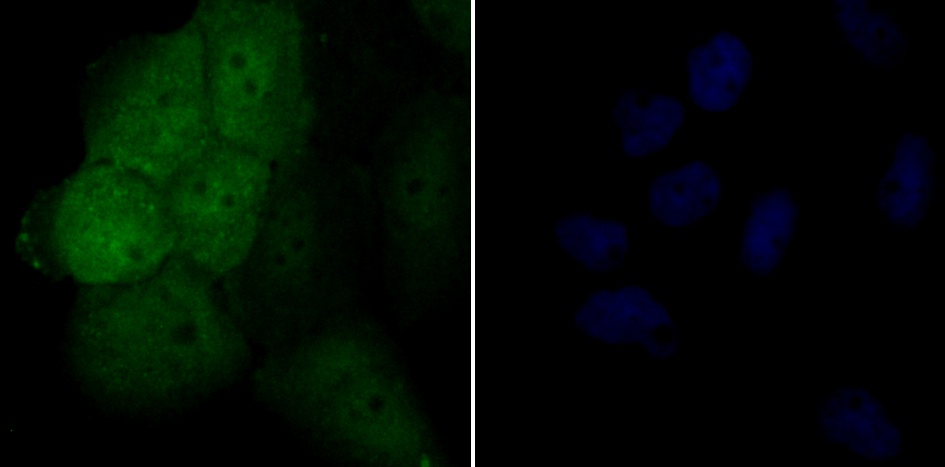 ICC staining Galectin 3 in A431 cells (green). Formalin fixed cells were permeabilized with 0.1% Triton X-100 in TBS for 10 minutes at room temperature and blocked with 1% Blocker BSA for 15 minutes at room temperature. Cells were probed with ER1803-82 at a dilution of 1:50 for 1 hour at room temperature, washed with PBS. Alexa Fluorc™ 488 Goat anti-Rabbit IgG was used as the secondary antibody at 1/100 dilution. The nuclear counter stain is DAPI (blue).
Bioworld Biotech only provide peptides for our antibodies and do not provide additional peptide customization services.
Price/Size :
USD 368/1mg/vial

Tips:
For phospho antibody, we provide phospho peptide(0.5mg) and non-phospho peptide(0.5mg).Describe :
Blocking peptides are peptides that bind specifically to the target antibody and block antibody binding. These peptide usually contains the epitope recognized by the antibody. Antibodies bound to the blocking peptide no longer bind to the epitope on the target protein. This mechanism is useful when non-specific binding is an issue, for example, in Western blotting (WB) and Immunohistochemistry (IHC). By comparing the staining from the blocked antibody versus the antibody alone, one can see which staining is specific; Specific binding will be absent from the western blot or IHC performed with the neutralized antibody.Formula:
Synthetic peptide was lyophilized with 100% acetonitrile and is supplied as a powder. Reconstitute with 0.1 ml DI water for a final concentration of 10 mg/ml.The purity is >90%,tested by HPLC and MS.
Storage:
The freeze-dried powder is more stable. For short time at 2-8°C. For long term storage store at -20°C.
Note :
This product is for research use only (RUO only). Not for use in diagnostic or therapeutic procedures.