
 DDB1 polyclonal antibody
  Datasheet
 COA
COA MSDS
MSDS SHIP
SHIP
Product Name :
DDB1 polyclonal antibody Background :
Damaged DNA binding protein (DDB) is a heterodimer composed of two subunits, p127 and p48, which are designated DDB1 and DDB2, respectively. The DDB heterodimer is involved in repairing DNA damaged by ultraviolet light. Specifically, DDB, also designated UV-damaged DNA binding protein (UV-DDB), xeroderma pigmentosum group E binding factor (XPE-BF) and hepatitis B virus X-associated protein 1 (XAP-1), binds to damaged cyclobutane pyrimidine dimers (CPDs). Mutations in the DDB2 gene are implicated as causes of xeroderma pigmentosum group E, an autosomal recessive disease in which patients are defective in nucleotide excision DNA repair. XPE is characterized by hypersensitivity of the skin to sunlight with a high frequency of skin cancer as well as neurologic abnormalities. The hepatitis B virus (HBV) X protein interacts with DDB1, which may mediate HBx transactivation. Product :
Rabbit IgG, 1mg/ml in PBS with 0.02% sodium azide, 50% glycerol, pH7.2 Storage&Stability :
Store at +4°C after thawing. Aliquot store at -20°C or -80°C. Avoid repeated freeze / thaw cycles. Specificity :
DDB1 polyclonal antibody detects endogenous levels of DDB1 protein. Immunogen :
Recombinant protein Conjugate :
Unconjugated Modification :
Unmodification
-
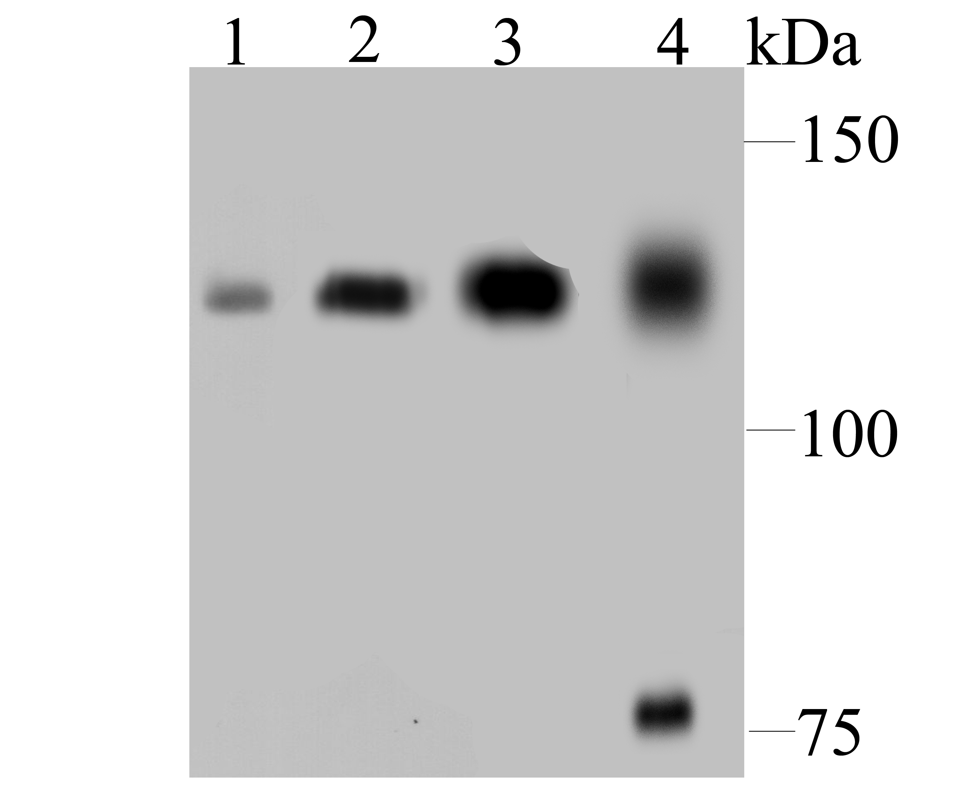 Western blot analysis of DDB1 on different lysates using anti-DDB1 antibody at 1/500 dilution. Positive control: Lane 1: HepG2 Lane 2: NIH-3T3 Lane 3: MCF-7 Lane 4: Rat kidney tissue
 -
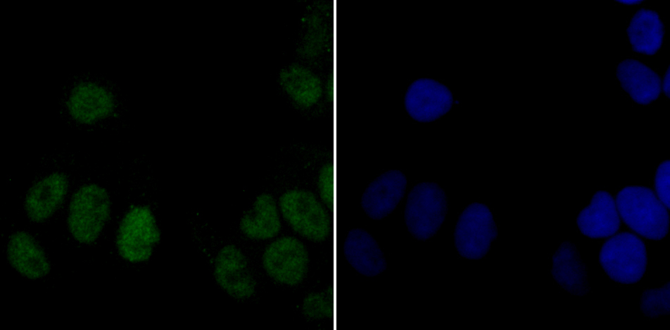 ICC staining DDB1 in Hela cells (green). The nuclear counter stain is DAPI (blue). Cells were fixed in paraformaldehyde, permeabilised with 0.25% Triton X100/PBS.
Bioworld Biotech only provide peptides for our antibodies and do not provide additional peptide customization services.
Price/Size :
USD 368/1mg/vial

Tips:
For phospho antibody, we provide phospho peptide(0.5mg) and non-phospho peptide(0.5mg).Describe :
Blocking peptides are peptides that bind specifically to the target antibody and block antibody binding. These peptide usually contains the epitope recognized by the antibody. Antibodies bound to the blocking peptide no longer bind to the epitope on the target protein. This mechanism is useful when non-specific binding is an issue, for example, in Western blotting (WB) and Immunohistochemistry (IHC). By comparing the staining from the blocked antibody versus the antibody alone, one can see which staining is specific; Specific binding will be absent from the western blot or IHC performed with the neutralized antibody.Formula:
Synthetic peptide was lyophilized with 100% acetonitrile and is supplied as a powder. Reconstitute with 0.1 ml DI water for a final concentration of 10 mg/ml.The purity is >90%,tested by HPLC and MS.
Storage:
The freeze-dried powder is more stable. For short time at 2-8°C. For long term storage store at -20°C.
Note :
This product is for research use only (RUO only). Not for use in diagnostic or therapeutic procedures.