
 AKT polyclonal antibody
  Datasheet
 COA
COA MSDS
MSDS SHIP
SHIP
Product Name :
AKT polyclonal antibody Background :
The serine/threonine kinase Akt family contains several members, including Akt1 (also designated PKB or RacPK), Akt2 (also designated PKBβ or RacPK-β) and Akt 3 (also designated PKBγ or thyoma viral proto-oncogene 3), which exhibit sequence homology with the protein kinase A and C families and are encoded by the c-Akt proto-oncogene. All members of the Akt family have a pleckstrin homology domain. Akt1 and Akt2 are activated by PDGF stimulation. This activation is dependent on PDGFR-β tyrosine residues 740 and 751, which bind the subunit of the phosphatidylinositol 3-kinase (PI 3-kinase) complex. Activation of Akt1 by insulin or insulin-growth factor-1(IGF-1) results in phosphorylation of both Thr 308 and Ser 473. Phosphorylation of both residues is important to generate a high level of Akt1 activity, and the phosphorylation of Thr 308 is not dependent on phosphorylation of Ser 473 in vivo. Thus, Akt proteins become phosphorylated and activated in insulin/IGF-1-stimulated cells by an upstream kinase(s). The activation of Akt1 and Akt2 is inhibited by the PI kinase inhibitor wortmannin, suggesting that the protein signals downstream of the PI kinases. Product :
Rabbit IgG, 1mg/ml in PBS with 0.02% sodium azide, 50% glycerol, pH7.2 Storage&Stability :
Store at +4°C after thawing. Aliquot store at -20°C or -80°C. Avoid repeated freeze / thaw cycles. Specificity :
AKT polyclonal antibody detects endogenous levels of AKT protein. Immunogen :
recombinant protein Conjugate :
Unconjugated Modification :
Unmodification
-
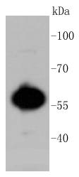 Western blot analysis of AKT1/2/3 on MCF-7 cell lysates using anti-AKT1/2/3 antibody at 1/1,000 dilution.
 -
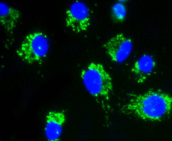 ICC staining AKT1/2/3 in A549 cells (green). The nuclear counter stain is DAPI (blue). Cells were fixed in paraformaldehyde, permeabilised with 0.25% Triton X100/PBS.
Ginseng root extract attenuates inflammation by inhibiting the MAPK/NF-κB signaling pathway and activating autophagy and p62-Nrf2-Keap1 signaling in vitro and in vivo
PMCID: Pubmed No.:34648903
siRNA directed against Annexin II receptor inhibits HeLa cell proliferation, migration and invasion and induces apoptosis via suppressing ERK1/2 and Akt signaling pathways
PMCID: Pubmed No.:31938090
Neuroprotective effect of Ginsenoside Re against neurotoxin‑induced Parkinson's disease models via induction of Nrf2
PMCID: Pubmed No.:35543148
Acteoside and ursolic acid synergistically protects H2O2-induced neurotrosis by regulation of AKT/mTOR signalling: from network pharmacology to experimental validation
PMCID: Pubmed No.:36102631
LncRNA BC promotes lung adenocarcinoma progression by modulating IMPAD1 alternative splicing
PMCID: Pubmed No.:36650118
γ-Glutamylcysteine rescues mice from TNBS-driven inflammatory bowel disease through regulating macrophages polarization
PMCID: Pubmed No.:36690783
A novel pharmaceutical preparation of Tripterygium wilfordii Hook. f. regulates macrophage polarization to alleviate inflammation in rheumatoid arthritis
PMCID: Pubmed No.:37738207
Bioworld Biotech only provide peptides for our antibodies and do not provide additional peptide customization services.
Price/Size :
USD 368/1mg/vial

Tips:
For phospho antibody, we provide phospho peptide(0.5mg) and non-phospho peptide(0.5mg).Describe :
Blocking peptides are peptides that bind specifically to the target antibody and block antibody binding. These peptide usually contains the epitope recognized by the antibody. Antibodies bound to the blocking peptide no longer bind to the epitope on the target protein. This mechanism is useful when non-specific binding is an issue, for example, in Western blotting (WB) and Immunohistochemistry (IHC). By comparing the staining from the blocked antibody versus the antibody alone, one can see which staining is specific; Specific binding will be absent from the western blot or IHC performed with the neutralized antibody.Formula:
Synthetic peptide was lyophilized with 100% acetonitrile and is supplied as a powder. Reconstitute with 0.1 ml DI water for a final concentration of 10 mg/ml.The purity is >90%,tested by HPLC and MS.
Storage:
The freeze-dried powder is more stable. For short time at 2-8°C. For long term storage store at -20°C.
Note :
This product is for research use only (RUO only). Not for use in diagnostic or therapeutic procedures.