
 ATP6V0A2 polyclonal antibody
  Datasheet
 COA
COA MSDS
MSDS SHIP
SHIP
Product Name :
ATP6V0A2 polyclonal antibody Background :
Vacuolar-type H+-ATPase (V-ATPase) is a multisubunit enzyme responsible for the acidification of eukaryotic intracellular organelles. V-ATPases pump protons against an electrochemical gradient, while F-ATPases reverse the process, thereby synthesizing ATP. A peripheral V1 domain, which is responsible for ATP hydrolysis, and an integral V0 domain, which is responsible for proton translocation, comprise the V-ATPase complex. Nine subunits (A–H) make up the V1 domain and five subunits (A, D, C, C' and C") make up the V0 domain. As part of the V0 domain, V-ATPase A2 (ATPase, H+ transporting, lysosomal V0 subunit a2), consists of 856 amino acids and is also known as ATP6V0A2, V-type proton ATPase subunit a isoform 2, vacuolar proton translocating ATPase subunit a isoform 2, lysosomal H(+)-transporting ATPase V0 subunit a2 or TJ6. V-ATPase A2 is a multi-pass membrane protein with localization in the cell membrane, endosome membrane and the subapical vesicles of the kidney’s proximal tubules. V-ATPase A2 plays an important role in Golgi function by regulating pH. Wrinkly skin syndrome (WSS) and cutis laxa type II (ARCL type II) are caused as a result of V-ATPase A2 defects. Product :
Rabbit IgG, 1mg/ml in PBS with 0.02% sodium azide, 50% glycerol, pH7.2 Storage&Stability :
Store at 4°C short term. Aliquot and store at -27°C long term. Avoid freeze-thaw cycles. Specificity :
ATP6V0A2 polyclonal antibody detects endogenous levels of ATP6V0A2 protein. Immunogen :
A synthetic peptide corresponding to residues in Human ATP6V0A2. Conjugate :
Unconjugated Modification :
Unmodification
-
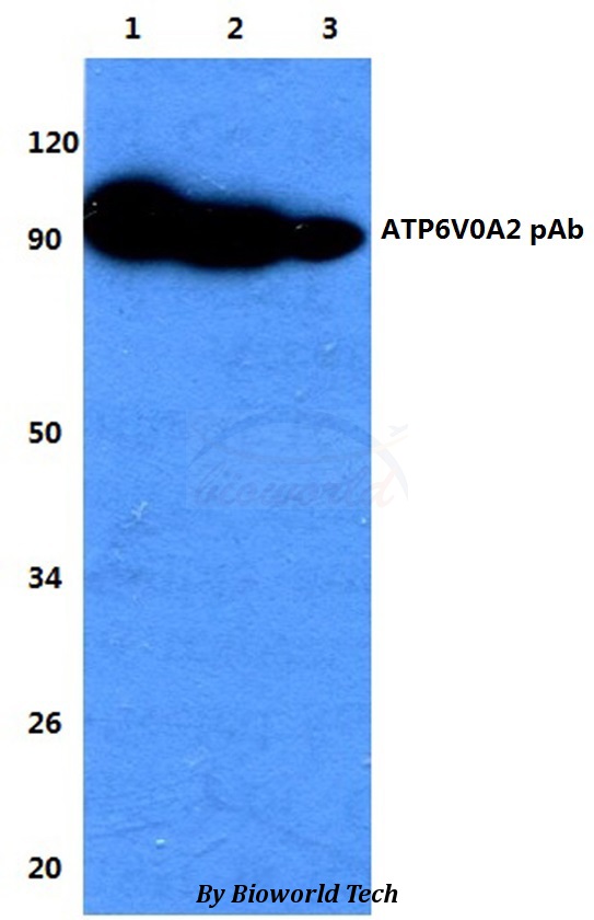 Western blot (WB) analysis of ATP6V0A2 polyclonal antibody at 1:500 dilution Lane1:HEK293T whole cell lysate Lane2:sp2/0 whole cell lysate Lane3:H9C9 whole cell lysate
Bioworld Biotech only provide peptides for our antibodies and do not provide additional peptide customization services.
Price/Size :
USD 368/1mg/vial

Tips:
For phospho antibody, we provide phospho peptide(0.5mg) and non-phospho peptide(0.5mg).Describe :
Blocking peptides are peptides that bind specifically to the target antibody and block antibody binding. These peptide usually contains the epitope recognized by the antibody. Antibodies bound to the blocking peptide no longer bind to the epitope on the target protein. This mechanism is useful when non-specific binding is an issue, for example, in Western blotting (WB) and Immunohistochemistry (IHC). By comparing the staining from the blocked antibody versus the antibody alone, one can see which staining is specific; Specific binding will be absent from the western blot or IHC performed with the neutralized antibody.Formula:
Synthetic peptide was lyophilized with 100% acetonitrile and is supplied as a powder. Reconstitute with 0.1 ml DI water for a final concentration of 10 mg/ml.The purity is >90%,tested by HPLC and MS.
Storage:
The freeze-dried powder is more stable. For short time at 2-8°C. For long term storage store at -20°C.
Note :
This product is for research use only (RUO only). Not for use in diagnostic or therapeutic procedures.