
 AKAP8 polyclonal antibody
  Datasheet
 COA
COA MSDS
MSDS SHIP
SHIP
Product Name :
AKAP8 polyclonal antibody Background :
The type II cAMP-protein kinase (PKA) is a multifunctional kinase with a broad range of substrates. Specificity of PKA signaling is thought to be mediated by the compartmentalization of the kinase to specific sites within the cell. To maintain this specific localization, the R subunit (RII) of PKA interacts with specific RII-anchoring proteins. The family of RII-anchoring proteins has been designated A-kinase anchoring proteins (AKAP). AKAP 95, also known as AKAP 8, is a nuclear matrix protein predominantly expressed in liver, heart, pancreas, kidney and skeletal muscle. During mitosis, AKAP 95 is recruited to the chromosomes and plays an essential role in mitotic progression. Characteristic of its family, AKAP 95 participates in PKA signaling through an interaction with the RII regulatory subunit. In addition, AKAP 95 forms a complex with HA95 and HDAC3 and is required for the deacetylation of Histone H3 in mitosis. Product :
Rabbit IgG, 1mg/ml in PBS with 0.02% sodium azide, 50% glycerol, pH7.2 Storage&Stability :
Store at 4°C short term. Aliquot and store at -20°C long term. Avoid freeze-thaw cycles. Specificity :
AKAP8 polyclonal antibody detects endogenous levels of AKAP8 protein. Immunogen :
Synthetic peptide, corresponding to amino acids 373-418 of Human AKAP8. Conjugate :
Unconjugated Modification :
Unmodification
-
 Western blot (WB) analysis of AKAP8 polyclonal antibody at 1:500 dilution Lane1:HepG2 cell lysate Lane2:Mouse kidney tissue lysate Lane3:Rat kidney tissue lysate
 -
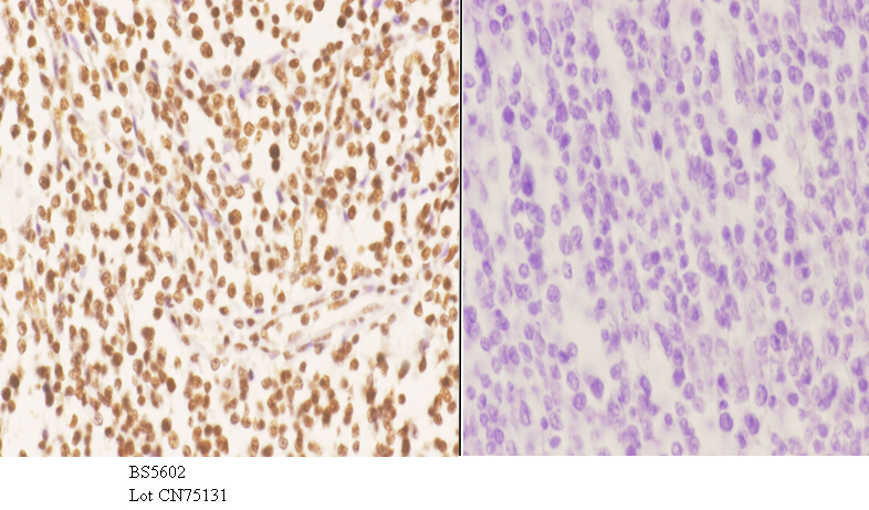 Immunohistochemistry (IHC) analyzes of AKAP8 pAb in paraffin-embedded human tonsil carcinoma tissue at 1:50.showing nucleus staining. Negative control (the right)Using PBS instead of primary antibody, secondary antibody is Goat Anti-Rabbit IgG-biotin followed by avidin-peroxidase.
 -
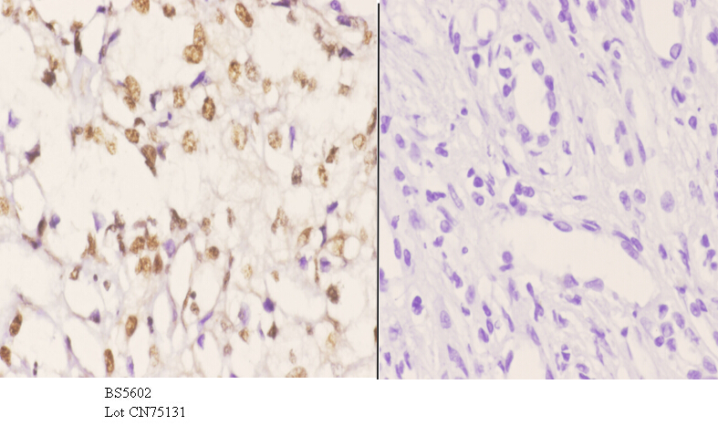 Immunohistochemistry (IHC) analyzes of AKAP8 pAb in paraffin-embedded human tonsil carcinoma tissue at 1:50.showing nucleus staining. Negative control (the right)Using PBS instead of primary antibody, secondary antibody is Goat Anti-Rabbit IgG-biotin followed by avidin-peroxidase.
Bioworld Biotech only provide peptides for our antibodies and do not provide additional peptide customization services.
Price/Size :
USD 368/1mg/vial

Tips:
For phospho antibody, we provide phospho peptide(0.5mg) and non-phospho peptide(0.5mg).Describe :
Blocking peptides are peptides that bind specifically to the target antibody and block antibody binding. These peptide usually contains the epitope recognized by the antibody. Antibodies bound to the blocking peptide no longer bind to the epitope on the target protein. This mechanism is useful when non-specific binding is an issue, for example, in Western blotting (WB) and Immunohistochemistry (IHC). By comparing the staining from the blocked antibody versus the antibody alone, one can see which staining is specific; Specific binding will be absent from the western blot or IHC performed with the neutralized antibody.Formula:
Synthetic peptide was lyophilized with 100% acetonitrile and is supplied as a powder. Reconstitute with 0.1 ml DI water for a final concentration of 10 mg/ml.The purity is >90%,tested by HPLC and MS.
Storage:
The freeze-dried powder is more stable. For short time at 2-8°C. For long term storage store at -20°C.
Note :
This product is for research use only (RUO only). Not for use in diagnostic or therapeutic procedures.