
 PKD2 (phospho-S876) polyclonal antibody
  Datasheet
 COA
COA MSDS
MSDS SHIP
SHIP
Product Name :
PKD2 (phospho-S876) polyclonal antibody Background :
Protein kinase D2 (PKD2) is one of three members of the protein kinase D family, including PKD1/PKCμ and PKD3/PKCν, that belong to the calcium/calmodulin superfamily of serine/threonine protein kinases. PKDs contain a conserved, carboxy-terminal catalytic domain, an amino-terminal regulatory region hallmarked by a PH domain that coordinates subcellular localization, and two zinc-finger/C1 lipid-binding domains that mediate activation of the enzyme in response to diacylglycerol (DAG) or phorbol ester. In addition to lipid-mediated activation, PKD catalytic activity can also be stimulated via phosphorylation of critical serine residues within the activation loop of the enzyme. Novel PKCs, such as PKCη and PKCε, have been shown to phosphorylate PKD1 at Ser744 and Ser748 (Ser706 and Ser710 in human PKD2), resulting in alleviation of autoinhibition of the enzyme mediated by PH domain interactions with the catalytic domain. Phosphorylation and activation of PKD isoforms has also been described for other upstream kinases. For example, casein kinase 2 (CK2) has been shown to phosphorylate PKD2 at Ser244, which promotes nuclear accumulation of PKD2, phosphorylation of HDAC7, and expression of Nur77. Although only a handfull of PKD2 effectors have been identified, PKD2 has been implicated in regulating an array of cellular events, including cell survival, development, growth, migration, and transformation. PKD2-mediated phosphorylation of at least one known substrate, phosphatidylinositol 4-kinase type IIIβ (PI4KIIIβ), also implicates PKD2 in the formation and regulation of exocytotic transport vesicles from the trans Golgi network. Product :
Rabbit IgG, 1mg/ml in PBS with 0.02% sodium azide, 50% glycerol, pH7.2 Storage&Stability :
Store at 4°C short term. Aliquot and store at -20°C long term. Avoid freeze-thaw cycles. Specificity :
PKD2 (phospho-S876) polyclonal antibody detects endogenous levels of PKD2 protein only when phosphorylated at Ser876. Immunogen :
Synthetic phosphopeptide derived from human PKD2 around the phosphorylation site of Serine 876 Conjugate :
Unconjugated Modification :
Phosphorylation
-
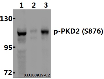 Western blot (WB) analysis of PKD2 (phospho-S876) polyclonal antibody at 1:500 dilution Lane1:DLD whole cell lysate(40µg) Lane2:MG63 whole cell lysate(40µg) Lane3:COS-7 whole cell lysate(40µg)
Bioworld Biotech only provide peptides for our antibodies and do not provide additional peptide customization services.
Price/Size :
USD 368/1mg/vial

Tips:
For phospho antibody, we provide phospho peptide(0.5mg) and non-phospho peptide(0.5mg).Describe :
Blocking peptides are peptides that bind specifically to the target antibody and block antibody binding. These peptide usually contains the epitope recognized by the antibody. Antibodies bound to the blocking peptide no longer bind to the epitope on the target protein. This mechanism is useful when non-specific binding is an issue, for example, in Western blotting (WB) and Immunohistochemistry (IHC). By comparing the staining from the blocked antibody versus the antibody alone, one can see which staining is specific; Specific binding will be absent from the western blot or IHC performed with the neutralized antibody.Formula:
Synthetic peptide was lyophilized with 100% acetonitrile and is supplied as a powder. Reconstitute with 0.1 ml DI water for a final concentration of 10 mg/ml.The purity is >90%,tested by HPLC and MS.
Storage:
The freeze-dried powder is more stable. For short time at 2-8°C. For long term storage store at -20°C.
Note :
This product is for research use only (RUO only). Not for use in diagnostic or therapeutic procedures.