
 Gα t1 (M104) polyclonal antibody
  Datasheet
 COA
COA MSDS
MSDS SHIP
SHIP
Product Name :
Gα t1 (M104) polyclonal antibody Background :
Heterotrimeric G proteins function to relay information from cell surface receptors to intracellular effectors. Each of a very broad range of receptors specifically detects an extracellular stimulus (a photon, pheromone, odorant, hormone or neurotransmitter) while the effectors (i.e. adenyl cyclase), which act to generate one or more intracellular messengers, are less numerous. In mammals, G protein α, β and γ polypeptides are encoded by at least 16, 4 and 7 genes, respectively. Most interest in G proteins has been focused on their α subunits, since these proteins bind and hydrolyze GTP and most obviously regulate the activity of the best studied effectors. Four distinct classes of Gα subunits have been identified; these include Gs, Gi, Gq and Gα 12/13. The Gi class comprises all the known α subunits that are susceptible to pertussis toxin modifications, including Gα i-1, Gα i-2, Gα i-3, Gα o, Gα t1, Gα t2, Gα z and Gα gust. In the well characterized visual system, photorhodopsin catalyzes the exchange of guanine nucleotides bound to the visual transducin Gα subunits (Gα t1 in rod cells and Gα t2 in cone cells). Product :
Rabbit IgG, 1mg/ml in PBS with 0.02% sodium azide, 50% glycerol, pH7.2 Storage&Stability :
Store at 4°C short term. Aliquot and store at -20°C long term. Avoid freeze-thaw cycles. Specificity :
Gα t1 (M104) polyclonal antibody detects endogenous levels of Gα t1 protein. Immunogen :
Synthetic peptide, corresponding to amino acids 71-120 of Human Gα t1. Conjugate :
Unconjugated Modification :
Unmodification
-
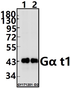 Western blot (WB) analysis of Gα t1 (M104) pAb at 1:500 dilution Lane1:The Eye tissue lysate of Mouse(20ug) Lane2:The Eye tissue lysate of Rat(20ug)
Bioworld Biotech only provide peptides for our antibodies and do not provide additional peptide customization services.
Price/Size :
USD 368/1mg/vial

Tips:
For phospho antibody, we provide phospho peptide(0.5mg) and non-phospho peptide(0.5mg).Describe :
Blocking peptides are peptides that bind specifically to the target antibody and block antibody binding. These peptide usually contains the epitope recognized by the antibody. Antibodies bound to the blocking peptide no longer bind to the epitope on the target protein. This mechanism is useful when non-specific binding is an issue, for example, in Western blotting (WB) and Immunohistochemistry (IHC). By comparing the staining from the blocked antibody versus the antibody alone, one can see which staining is specific; Specific binding will be absent from the western blot or IHC performed with the neutralized antibody.Formula:
Synthetic peptide was lyophilized with 100% acetonitrile and is supplied as a powder. Reconstitute with 0.1 ml DI water for a final concentration of 10 mg/ml.The purity is >90%,tested by HPLC and MS.
Storage:
The freeze-dried powder is more stable. For short time at 2-8°C. For long term storage store at -20°C.
Note :
This product is for research use only (RUO only). Not for use in diagnostic or therapeutic procedures.