
 DRP-2 (P510) polyclonal antibody
  Datasheet
 COA
COA MSDS
MSDS SHIP
SHIP
Product Name :
DRP-2 (P510) polyclonal antibody Background :
Dystrophin, utrophin and dystrophin-related protein 2 (DRP2) are Actin-binding proteins that are involved in anchoring the cytoskeleton to the plasma membrane. Dystrophin is the protein product of the Duchenne/Becker muscular dystrophy gene. Dystrophin is expressed in muscle and brain tissues, where it is localized to the inner surface of the plasma membrane. Evidence suggests that the upregulation of utrophin (also known as DRP1) can reduce the dystrophic pathology. DRP2 is principally expressed in the brain and spinal cord. Analysis of DRP2 expression in rat brain on SDS-PAGE reveals a characteristic quartet of bands from 100-120 kDa. DRP2 exhibits a punctate staining pattern of rat neuronal dendrites and in neuropil. DRP2 forms a complex with dystroglycan at the surface of myelin-forming Schwann cells and may play a role in the terminal stages of myelinogenesis in the peripheral nervous system. The gene encoding human DRP2 maps to chromosome Xq22. Product :
Rabbit IgG, 1mg/ml in PBS with 0.02% sodium azide, 50% glycerol, pH7.2 Storage&Stability :
Store at 4°C short term. Aliquot and store at -20°C long term. Avoid freeze-thaw cycles. Specificity :
DRP-2 (P510) polyclonal antibody detects endogenous levels of DRP-2 protein. Immunogen :
Synthetic peptide, corresponding to amino acids 468-532 of Human DRP-2. Conjugate :
Unconjugated Modification :
Unmodification
-
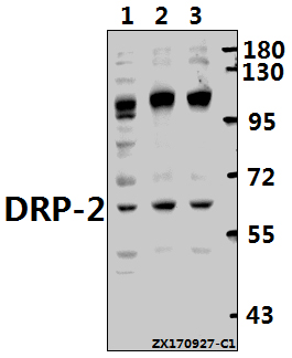 Western blot (WB) analysis of DRP-2 (P510) pAb at 1:500 dilution Lane1:H1792 whole cell lysate(40ug) Lane2:SK-OVCAR3 whole cell lysate(40ug) Lane3:A549 whole cell lysate(40ug)
 -
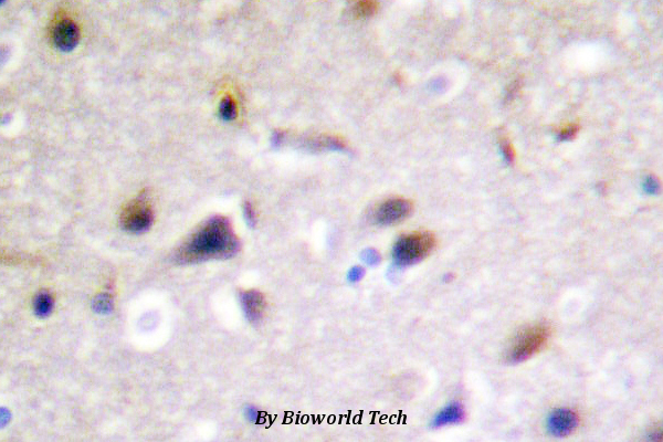 Immunohistochemistry (IHC) analyzes of DRP-2 (P510) pAb in paraffin-embedded human brain tissue .
Proteomics Reveals Intersexual Differences in the Rat Brain Hippocampus
PMCID: Pubmed No.:23381953
Proteomic Changes in Female Rat Hippocampus Following Exposure to a Terrified Sound Stress
PMCID: Pubmed No.:24510750
Proteomic Changes in Female Rat Hippocampus Following Exposure to a Terrified Sound Stress
PMCID: Pubmed No.:24510750
Proteomic Analysis of Protein Expression Throughout Disease Progression in a Mouse Model of Alzheimer’s Disease
PMCID: Pubmed No.:26401771
Bioworld Biotech only provide peptides for our antibodies and do not provide additional peptide customization services.
Price/Size :
USD 368/1mg/vial

Tips:
For phospho antibody, we provide phospho peptide(0.5mg) and non-phospho peptide(0.5mg).Describe :
Blocking peptides are peptides that bind specifically to the target antibody and block antibody binding. These peptide usually contains the epitope recognized by the antibody. Antibodies bound to the blocking peptide no longer bind to the epitope on the target protein. This mechanism is useful when non-specific binding is an issue, for example, in Western blotting (WB) and Immunohistochemistry (IHC). By comparing the staining from the blocked antibody versus the antibody alone, one can see which staining is specific; Specific binding will be absent from the western blot or IHC performed with the neutralized antibody.Formula:
Synthetic peptide was lyophilized with 100% acetonitrile and is supplied as a powder. Reconstitute with 0.1 ml DI water for a final concentration of 10 mg/ml.The purity is >90%,tested by HPLC and MS.
Storage:
The freeze-dried powder is more stable. For short time at 2-8°C. For long term storage store at -20°C.
Note :
This product is for research use only (RUO only). Not for use in diagnostic or therapeutic procedures.