
 AKT1 (P125) polyclonal antibody
  Datasheet
 COA
COA MSDS
MSDS SHIP
SHIP
Product Name :
AKT1 (P125) polyclonal antibody Background :
AKT, also known as protein kinase B (PKB), is a 57 kDa serine/threonine protein kinase. There are three mammalian isoforms of Akt: AKT1 (PKB alpha), AKT2 (PKB beta) and AKT3 (PKB gamma) with AKT2 and AKT3 being approximately 82% identical with the AKT1 isoform. Each isoform has a pleckstrin homology (PH) domain, a kinase domain and a carboxy terminal regulatory domain. AKT was originally cloned from the retrovirus AKT8, and is a key regulator of many signal transduction pathways. Its tight control over cell proliferation and cell viability are manifold; overexpression or inappropriate activation of AKT has been seen in many types of cancer. Product :
Rabbit IgG, 1mg/ml in PBS with 0.02% sodium azide, 50% glycerol, pH7.2 Storage&Stability :
Store at 4°C short term. Aliquot and store at -20°C long term. Avoid freeze-thaw cycles. Specificity :
AKT1 (P125) polyclonal antibody detects endogenous levels of AKT1 protein, this antibody does not cross-react with AKT2 or AKT3. Immunogen :
Synthetic peptide, corresponding to amino acids 100-150 of Human AKT1. Conjugate :
Unconjugated Modification :
Unmodification
-
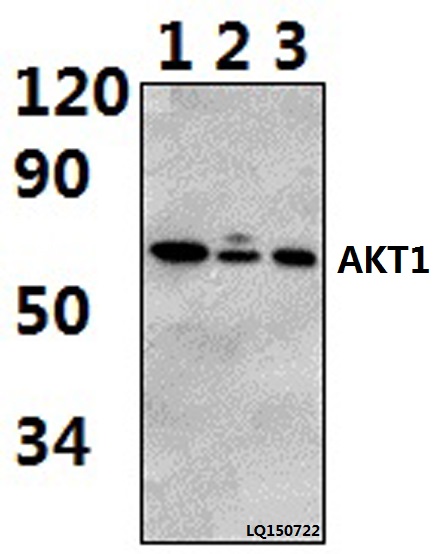 Western blot (WB) analysis of AKT1 (P125) polyclonal antibody at 1:500 dilution Lane1:A549 whole cell lysate(40ug) Lane2:NIH-3T3 whole cell lysate(40ug) Lane3:PC12 whole cell lysate(40ug)
 -
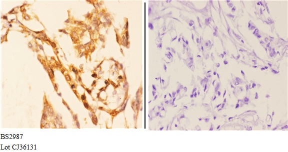 Immunohistochemistry (IHC) analyzes of AKT1 (P125) pAb in paraffin-embedded human breast carcinoma tissue at 1:50.showing Cytoplasm, Nucleus and Cell membrane staining. Negative control (the right)Using PBS instead of primary antibody, secondary antibody is Goat Anti-Rabbit IgG-biotin followed by avidin-peroxidase.
 -
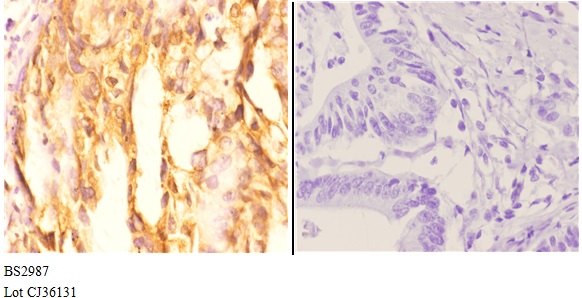 Immunohistochemistry (IHC) analyzes of AKT1 (P125) pAb in paraffin-embedded human breast carcinoma tissue at 1:50.showing Cytoplasm, Nucleus and Cell membrane staining. Negative control (the right)Using PBS instead of primary antibody, secondary antibody is Goat Anti-Rabbit IgG-biotin followed by avidin-peroxidase.
 -
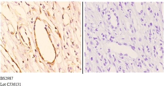 Immunohistochemistry (IHC) analyzes of AKT1 (P125) pAb in paraffin-embedded human breast carcinoma tissue at 1:50.showing Cytoplasm, Nucleus and Cell membrane staining. Negative control (the right)Using PBS instead of primary antibody, secondary antibody is Goat Anti-Rabbit IgG-biotin followed by avidin-peroxidase.
Luteolin inhibits inflammatory response and improves insulin sensitivity in the endothelium
PMCID: Pubmed No.:21081149
Modified Si-Miao-San Extract inhibits inflammatory response and modulates insulin sensitivity in hepatocytes through an IKKβ/IRS-1/Akt-dependent pathway
PMCID: Pubmed No.:21296137
Prevention of FGF-2-induced angiogenesis by scopoletin, a coumarin compound isolated from Erycibe obtusifolia Benth, and its mechanism of action
PMCID: Pubmed No.:21896339
Anti-inflammatory and immunomodulatory mechanisms of artemisinin on contact hypersensitivity
PMCID: Pubmed No.:22122827
Cellular FLICE-inhibitory protein protects against cardiac remodelling after myocardial infarction
PMCID: Pubmed No.:22202974
Puerarin attenuates endothelial insulin resistance through inhibition of inflammatory response in an IKKβ/IRS-1-dependent manner
PMCID: Pubmed No.:22314193
Interferon regulatory factor 3 is a negative regulator of pathological cardiac hypertrophy
PMCID: Pubmed No.:23307144
Small interfering RNA targeting ILK inhibits metastasis in human tongue cancer cells through repression of epithelial-to-mesenchymal transition
PMCID: Pubmed No.:23707970
Mindin regulates vascular smooth muscle cell phenotype and prevents neointima formation
PMCID: Pubmed No.:25751394
Arginine ADP-ribosyltransferase 1 promotes angiogenesis in colorectal cancer via the PI3K/Akt pathway
PMCID: Pubmed No.:26847718
Downregulation of Foxc2 enhances apoptosis induced by 5-fluorouracil through activation of MAPK and AKT pathways in colorectal cancer
PMCID: Pubmed No.:26893778
Lentivirus-induced knockdown of LRP1 induces osteoarthritic-like effects and increases susceptibility to apoptosis in chondrocytes via the nuclear factor-κB pathway
PMCID: Pubmed No.:26170918
To Explore the Pathogenesis of Vascular Lesion of Type 2 Diabetes Mellitus Based on the PI3K/Akt Signaling Pathway
PMCID: Pubmed No.:31179340
Arginine ADP-ribosyltransferase 1 Regulates Glycolysis in Colorectal Cancer via the PI3K/AKT/HIF1α Pathway
PMCID: Pubmed No.:35798928
CDK2 and CDK4 targeted liensinine inhibits the growth of bladder cancer T24 cells
PMCID: Pubmed No.:37423554
Krüppel-like Factor 12 regulates aging ovarian granulosa cell apoptosis by repressing SPHK1 transcription and Sphingosine-1-phosphate (S1P) production
PMCID: Pubmed No.:37543362
Resveratrol improves palmitic acid‑induced insulin resistance via the DDIT4/mTOR pathway in C2C12 cells
PMCID: Pubmed No.:37594055
Bioworld Biotech only provide peptides for our antibodies and do not provide additional peptide customization services.
Price/Size :
USD 368/1mg/vial

Tips:
For phospho antibody, we provide phospho peptide(0.5mg) and non-phospho peptide(0.5mg).Describe :
Blocking peptides are peptides that bind specifically to the target antibody and block antibody binding. These peptide usually contains the epitope recognized by the antibody. Antibodies bound to the blocking peptide no longer bind to the epitope on the target protein. This mechanism is useful when non-specific binding is an issue, for example, in Western blotting (WB) and Immunohistochemistry (IHC). By comparing the staining from the blocked antibody versus the antibody alone, one can see which staining is specific; Specific binding will be absent from the western blot or IHC performed with the neutralized antibody.Formula:
Synthetic peptide was lyophilized with 100% acetonitrile and is supplied as a powder. Reconstitute with 0.1 ml DI water for a final concentration of 10 mg/ml.The purity is >90%,tested by HPLC and MS.
Storage:
The freeze-dried powder is more stable. For short time at 2-8°C. For long term storage store at -20°C.
Note :
This product is for research use only (RUO only). Not for use in diagnostic or therapeutic procedures.