
 PU.1 (R212) polyclonal antibody
  Datasheet
 COA
COA MSDS
MSDS SHIP
SHIP
Product Name :
PU.1 (R212) polyclonal antibody Background :
The Ets transcription factor family (Ets-1, Ets-2, Erg-1–3, Elk-1, Elf-1, Elf-5, NERF, PU.1, PEA3, ERM, FEV, ER8l, Fli-1, TEL, Spi-B, ESE-1, ESE-3A, Net, ABT1 and ERF) are DNA-binding proteins that influence lymphoid development and activity. The Ets family monomeric proteins bind the consensus DNA site GGA(A/T) through a unique winged helix-turn-helix motif known as the Ets domain. PU.1 (Spi-1/Spi-A), Spi-B and Spi-C are closely related Ets family members which share a conserved divergent sequence within the Ets domain that enables their binding to the non-canonical AGAA sites. PU.1 transactivates a large number of B cell genes, such as those encoding CD72, CD20 and Btk, and Spi-B enhances expression of many of these same target genes. PU.1 is expressed in a wide variety of hematopoetic cells, including B cells, early T cells, megakaryocytes, granulocytes, mast cells, immature erythrocytes and myeloid cells. Alternatively, Spi-B expression is limited to B cells and immature T cells, where expression accumulates through T lineage commitment and then is dramatically absent following the β selection checkpoint. Product :
Rabbit IgG, 1mg/ml in PBS with 0.02% sodium azide, 50% glycerol, pH7.2 Storage&Stability :
Store at 4°C short term. Aliquot and store at -20°C long term. Avoid freeze-thaw cycles. Specificity :
PU.1 (R212) polyclonal antibody detects endogenous levels of PU.1 protein. Immunogen :
Synthetic peptide, corresponding to amino acids 180-230 of Human PU.1. Conjugate :
Unconjugated Modification :
Unmodification
-
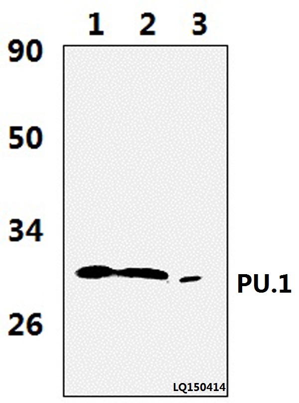 Western blot (WB) analysis of PU.1 (R212) polyclonal antibody at 1:1000 dilution Lane1:JURKAT whole cell lysate(37ug) Lane2:THP-1 whole cell lysate(37ug) Lane3:The blood cell tissue lysate of Rat(36ug)
 -
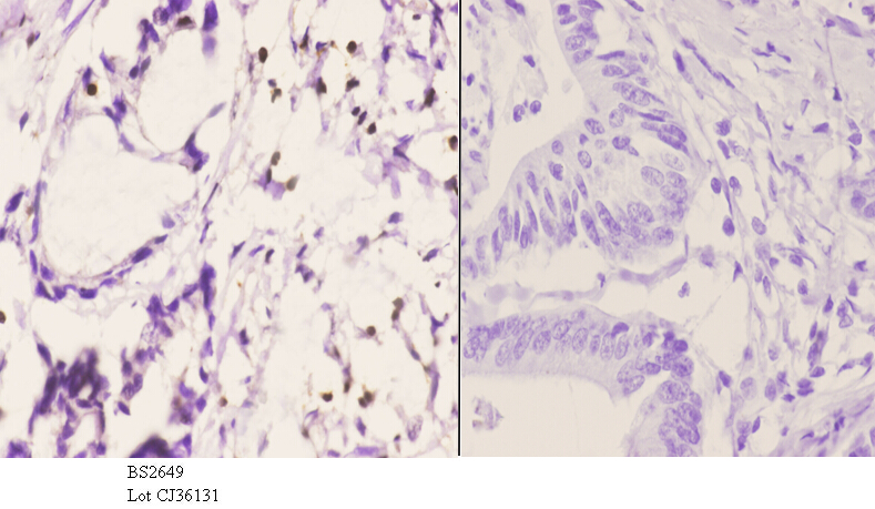 Immunohistochemistry (IHC) analyzes of PU.1 (R212) pAb in paraffin-embedded human colon carcinoma tissue at 1:50.showing nucleus staining. Negative control (the right)Using PBS instead of primary antibody, secondary antibody is Goat Anti-Rabbit IgG-biotin followed by avidin-peroxidase.
 -
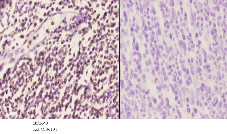 Immunohistochemistry (IHC) analyzes of PU.1 (R212) pAb in paraffin-embedded human colon carcinoma tissue at 1:50.showing nucleus staining. Negative control (the right)Using PBS instead of primary antibody, secondary antibody is Goat Anti-Rabbit IgG-biotin followed by avidin-peroxidase.
 -
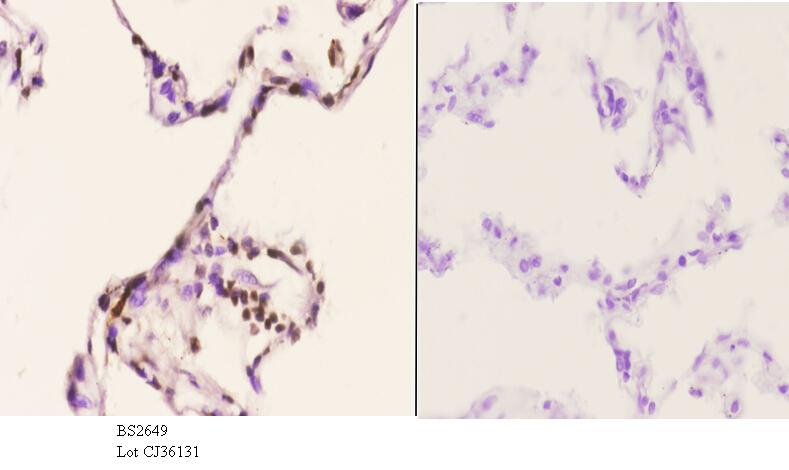 Immunohistochemistry (IHC) analyzes of PU.1 (R212) pAb in paraffin-embedded human colon carcinoma tissue at 1:50.showing nucleus staining. Negative control (the right)Using PBS instead of primary antibody, secondary antibody is Goat Anti-Rabbit IgG-biotin followed by avidin-peroxidase.
TanshinoneIIA ameliorates inflammatory microenvironment of colon cancer cells via repression of microRNA-155
PMCID: Pubmed No.:22982040
Calcitonin Gene-Related Peptide and Cyclic Adenosine 5'-Monophosphate/Protein Kinase A Pathway Promote IL-9 Production in Th9 Differentiation Process.
PMCID: Pubmed No.:23509367
TL1A modulates the severity of colitis by promoting Th9 differentiation and IL-9 secretion
PMCID: Pubmed No.:31176785
Th9 cytokines curb cervical cancer progression and immune evasion
PMCID: Pubmed No.:31563404
Bioworld Biotech only provide peptides for our antibodies and do not provide additional peptide customization services.
Price/Size :
USD 368/1mg/vial

Tips:
For phospho antibody, we provide phospho peptide(0.5mg) and non-phospho peptide(0.5mg).Describe :
Blocking peptides are peptides that bind specifically to the target antibody and block antibody binding. These peptide usually contains the epitope recognized by the antibody. Antibodies bound to the blocking peptide no longer bind to the epitope on the target protein. This mechanism is useful when non-specific binding is an issue, for example, in Western blotting (WB) and Immunohistochemistry (IHC). By comparing the staining from the blocked antibody versus the antibody alone, one can see which staining is specific; Specific binding will be absent from the western blot or IHC performed with the neutralized antibody.Formula:
Synthetic peptide was lyophilized with 100% acetonitrile and is supplied as a powder. Reconstitute with 0.1 ml DI water for a final concentration of 10 mg/ml.The purity is >90%,tested by HPLC and MS.
Storage:
The freeze-dried powder is more stable. For short time at 2-8°C. For long term storage store at -20°C.
Note :
This product is for research use only (RUO only). Not for use in diagnostic or therapeutic procedures.