
 ERAP1 (K467) polyclonal antibody
  Datasheet
 COA
COA MSDS
MSDS SHIP
SHIP
Product Name :
ERAP1 (K467) polyclonal antibody Background :
The endoplasmic reticulum (ER) aminopeptidase 1 (ERAP1) is a 120 kDa protein localized to the lumen of the ER, which removes NH2-terminal residues from many antigenic precursors for MHC class I peptide presentation. Peptides that are presented by MHC class I on the surface of a cell must be 8-11 residues long, and ERAP1 specifically trims peptides of 9 amino acids or more. ERAP1 is also induced by interferon-γ. The gene encoding human ERAP1 maps to chromosome 5q15. ERAP1 has previously been characterized as adipocyte-derived leucine aminopeptidase (A-LAP), puromycin-insensitive leucine-specific aminopeptidase (PILS-AP) and aminopeptidase regulator of TNFR1 shedding (ARTS-1). A-LAP is thought to inactivate several bioactive peptides, including angiotensin II and, subsequently, may be involved in the regulation of blood pressure. PILS-AP is described as playing a role in angiogenesis by regulating the proliferation and migration of endothelial cells, and ARTS-1 is characterized as a TNFR1 binding protein that promotes TNFR1 shedding. Further research will be necessary to fully elucidate the functions of this protein. Product :
Rabbit IgG, 1mg/ml in PBS with 0.02% sodium azide, 50% glycerol, pH7.2 Storage&Stability :
Store at 4°C short term. Aliquot and store at -20°C long term. Avoid freeze-thaw cycles. Specificity :
ERAP1 (K467) polyclonal antibody detects endogenous levels of ERAP1 protein. Immunogen :
Synthetic peptide, corresponding to amino acids 435-489 of Human ERAP1. Conjugate :
Unconjugated Modification :
Unmodification
-
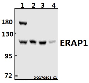 Western blot (WB) analysis of ERAP1 (K467) pAb at 1:500 dilution Lane1:HepG2 whole cell lysate(40ug) Lane2:HEK293T whole cell lysate(20ug) Lane3:PC12 whole cell lysate(20ug) Lane4:AML-12 whole cell lysate(40ug)
 -
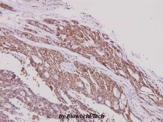 Immunohistochemistry (IHC) analyzes of ERAP1 (K467) pAb in paraffin-embedded human breast carcinoma tissue at 1:100.
Bioworld Biotech only provide peptides for our antibodies and do not provide additional peptide customization services.
Price/Size :
USD 368/1mg/vial

Tips:
For phospho antibody, we provide phospho peptide(0.5mg) and non-phospho peptide(0.5mg).Describe :
Blocking peptides are peptides that bind specifically to the target antibody and block antibody binding. These peptide usually contains the epitope recognized by the antibody. Antibodies bound to the blocking peptide no longer bind to the epitope on the target protein. This mechanism is useful when non-specific binding is an issue, for example, in Western blotting (WB) and Immunohistochemistry (IHC). By comparing the staining from the blocked antibody versus the antibody alone, one can see which staining is specific; Specific binding will be absent from the western blot or IHC performed with the neutralized antibody.Formula:
Synthetic peptide was lyophilized with 100% acetonitrile and is supplied as a powder. Reconstitute with 0.1 ml DI water for a final concentration of 10 mg/ml.The purity is >90%,tested by HPLC and MS.
Storage:
The freeze-dried powder is more stable. For short time at 2-8°C. For long term storage store at -20°C.
Note :
This product is for research use only (RUO only). Not for use in diagnostic or therapeutic procedures.