
 DGK-δ (S66) polyclonal antibody
  Datasheet
 COA
COA MSDS
MSDS SHIP
SHIP
Product Name :
DGK-δ (S66) polyclonal antibody Background :
Diacylglycerol kinases (DGKs) phosphorylate diacylglycerol (DAG) to produce phosphatidic acid. DAG and phosphatidic acid are lipids that act as second messengers in signaling cascades. DGK-α influences cell activation and secretion of lethal exosomes, which in turn control cell death. DGK-β is abundant in restricted brain regions such as the caudate putamen and olfactory tubercle. DGK-γ encodes full-length and truncated transcripts that are present in a range of human tissues, with greatest expression observed in retina. DGK-δ is most abundant in skeletal muscle. DGK-ε shows specificity for arachidonylcontaining diacylglycerol and is expressed predominantly in testis. DGK-θ is most abundant in the cerebellum and hippocampus. DGK-ι is present in brain and retina as a predominant transcript of more than 12 kb, including a long 3-prime untranslated region, with additional low abundance transcripts of 9.5 and 7.5 kb. DGK-η is closely related to DGK-δ. DGK-ζ is most abundant in brain and muscle. DGKs have structural motifs that play regulatory roles, and these motifs form the basis for dividing the DGKs into five subtypes. Product :
Rabbit IgG, 1mg/ml in PBS with 0.02% sodium azide, 50% glycerol, pH7.2 Storage&Stability :
Store at 4°C short term. Aliquot and store at -20°C long term. Avoid freeze-thaw cycles. Specificity :
DGK-δ (S66) polyclonal antibody detects endogenous levels of DGK-δ protein. Immunogen :
Synthetic peptide, corresponding to amino acids 34-88 of Human DGK-δ. Conjugate :
Unconjugated Modification :
Unmodification
-
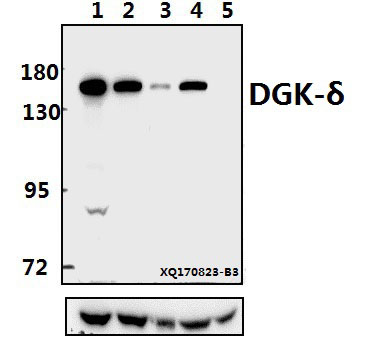 Western blot (WB) analysis of DGK-δ (S66) pAb at 1:500 dilution Lane1:A549 whole cell lysate(40ug) Lane2:Hela whole cell lysate(40ug) Lane3:A2780 whole cell lysate(40ug) Lane4:PC12 whole cell lysate(40ug) Lane5:3T3-L1 whole cell lysate(40ug)
 -
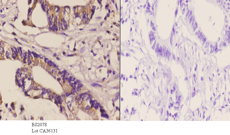 Immunohistochemistry (IHC) analyzes of DGK-δ (S66) pAb in paraffin-embedded human Rectum carcinoma tissue at 1:50.showing cytoplasmic staining. Negative control (the right)Using PBS instead of primary antibody, secondary antibody is Goat Anti-Rabbit IgG-biotin followed by avidin-peroxidase.
Bioworld Biotech only provide peptides for our antibodies and do not provide additional peptide customization services.
Price/Size :
USD 368/1mg/vial

Tips:
For phospho antibody, we provide phospho peptide(0.5mg) and non-phospho peptide(0.5mg).Describe :
Blocking peptides are peptides that bind specifically to the target antibody and block antibody binding. These peptide usually contains the epitope recognized by the antibody. Antibodies bound to the blocking peptide no longer bind to the epitope on the target protein. This mechanism is useful when non-specific binding is an issue, for example, in Western blotting (WB) and Immunohistochemistry (IHC). By comparing the staining from the blocked antibody versus the antibody alone, one can see which staining is specific; Specific binding will be absent from the western blot or IHC performed with the neutralized antibody.Formula:
Synthetic peptide was lyophilized with 100% acetonitrile and is supplied as a powder. Reconstitute with 0.1 ml DI water for a final concentration of 10 mg/ml.The purity is >90%,tested by HPLC and MS.
Storage:
The freeze-dried powder is more stable. For short time at 2-8°C. For long term storage store at -20°C.
Note :
This product is for research use only (RUO only). Not for use in diagnostic or therapeutic procedures.