
 TYR (R501) polyclonal antibody
  Datasheet
 COA
COA MSDS
MSDS SHIP
SHIP
Product Name :
TYR (R501) polyclonal antibody Background :
Tyrosinase (TYR), a type I membrane protein and copper-containing enzyme, is involved in the production of melanin, the primary pigment found in vertebrates. Melanin biogenesis requires the enzymatic activity of TYR, which catalyzes the critical and rate-limiting step of tyrosine hydroxylation in the biosynthesis of melanin. Defects effecting TYR activity result in various forms of albinism. The TYR-related proteins, TRP1 and TRP2, are also specifically expressed in melanocytes, and they likewise contribute to the synthesis of melanin within the melanosomes. The TRPs, including TYR, all share a similar transmembrane region, contain two metal-binding regions and a cysteinerich epidermal growth factor motif, and are localized in the melanosomal membrane. These proteins, however, have distinct catalytic activity, and they individually contribute to the biosynthesis of melanin biopolymers. The TRPs are believed to exists as a multi-enzyme complex, as these proteins form aggregates together, and the expression of TRP1 also helps stabilize TYR in melanocytes. Product :
Rabbit IgG, 1mg/ml in PBS with 0.02% sodium azide, 50% glycerol, pH7.2 Storage&Stability :
Store at 4°C short term. Aliquot and store at -20°C long term. Avoid freeze-thaw cycles. Specificity :
Tyrosinase (R501) polyclonal antibody detects endogenous levels of Tyrosinase protein. Immunogen :
Synthetic peptide, corresponding to amino acids 470-520 of Human TYR. Conjugate :
Unconjugated Modification :
Unmodification
-
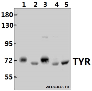 Western blot (WB) analysis of TYR (R501) pAb at 1:500 dilution Lane1:A375 whole cell lysate(40ug) Lane2:Podocyte whole cell lysate(40ug) Lane3:Beas-2B whole cell lysate(40ug) Lane4:PMVEC whole cell lysate(40ug) Lane5:BV2 whole cell lysate(40ug)
 -
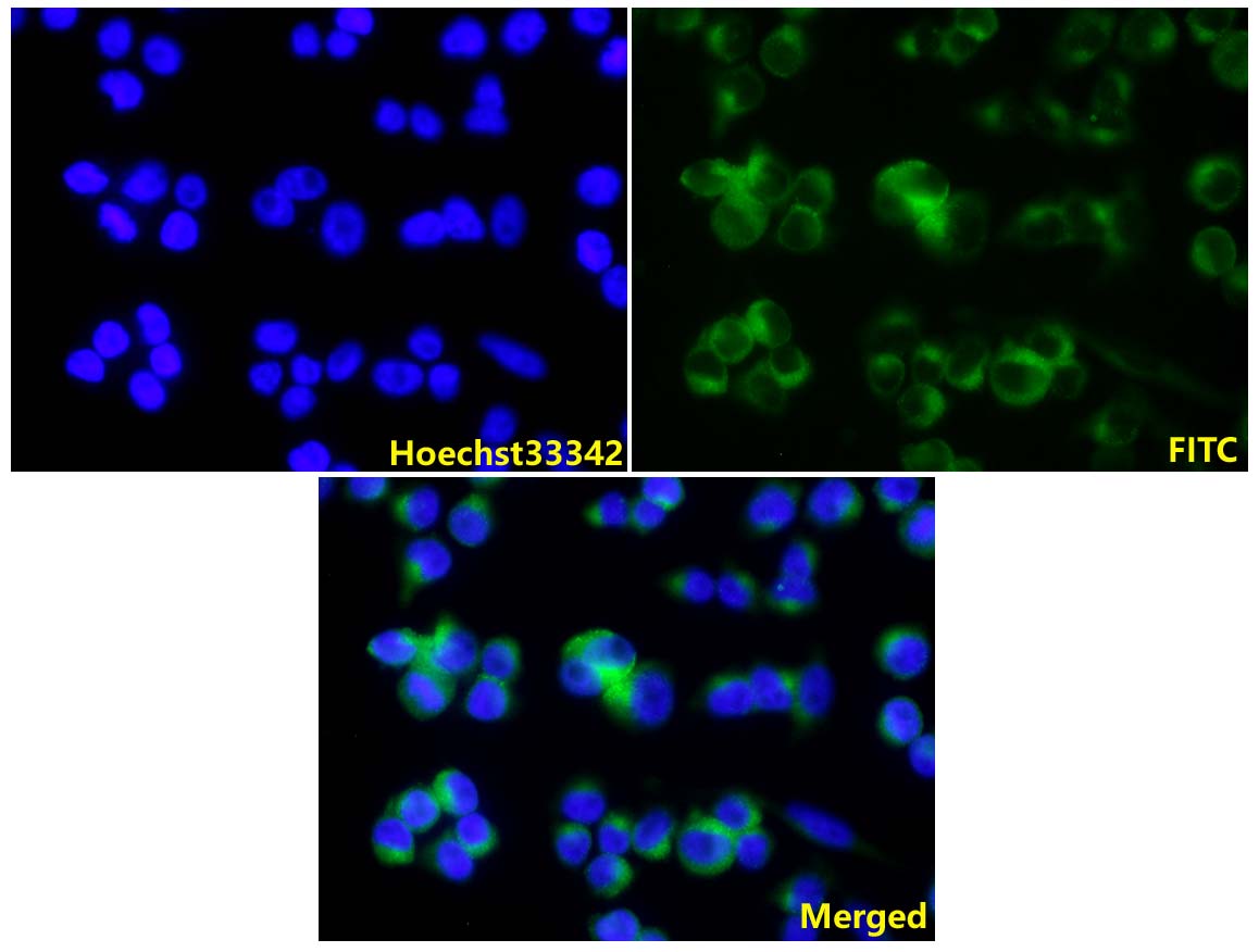 IF image of BS1484 stained A375 cells.The cells were 4% paraformaldehyde fixed (20 min) and then incubated in 10% normal goat serum for 1h to permeabilise the cells and block non-specific protein-protein interactions. The cells were then incubated with the antibody TYR (R501) pAb #BS1484(1:100) at 10µg/ml overnight at +4°C. The secondary antibody (Green) was Goat Anti-Mouse IgG (H+L) FITC#BS10950 used at a 1/400 dilution for 2h. Hoechst33342 #BD5011 was used to stain the cell nuclei (blue).
Flavonoids, apigenin and icariin exert potent melanogenic activities in murine B16 melanoma cells
PMCID: Pubmed No.:20638260
Involvement of p38 MAPK signaling pathway in the anti-melanogenic effect of San-bai-tang, a Chinese herbal formula, in B16 cells
PMCID: Pubmed No.:20837127
Wnt10b Promotes Differentiation of Mouse Hair Follicle Melanocytes
PMCID: Pubmed No.:23569433
SCF/c-kit signaling is required in 12-O-tetradecanoylphorbol-13-acetate-induced migration and differentiation of hair follicle melanocytes for epidermal pigmentation
PMCID: Pubmed No.:25727244
Identification of Sitogluside as a Potential Skin-Pigmentation-Reducing Agent through Network Pharmacology
PMCID: Pubmed No.:34603597
Fuzhuan Brick Tea Boosts Melanogenesis and Prevents Hair Graying through Reduction of Oxidative Stress via NRF2-HO-1 Signaling
PMCID: Pubmed No.:35326249
Establishment and validation of evaluation models for post-inflammatory pigmentation abnormalities
PMCID: Pubmed No.:36389813
Pueraria protein extract inhibits melanogenesis and promotes melanoma cell apoptosis through the regulation of MITF and mitochondrial‑related pathways
PMCID: Pubmed No.:36734267
Bioworld Biotech only provide peptides for our antibodies and do not provide additional peptide customization services.
Price/Size :
USD 368/1mg/vial

Tips:
For phospho antibody, we provide phospho peptide(0.5mg) and non-phospho peptide(0.5mg).Describe :
Blocking peptides are peptides that bind specifically to the target antibody and block antibody binding. These peptide usually contains the epitope recognized by the antibody. Antibodies bound to the blocking peptide no longer bind to the epitope on the target protein. This mechanism is useful when non-specific binding is an issue, for example, in Western blotting (WB) and Immunohistochemistry (IHC). By comparing the staining from the blocked antibody versus the antibody alone, one can see which staining is specific; Specific binding will be absent from the western blot or IHC performed with the neutralized antibody.Formula:
Synthetic peptide was lyophilized with 100% acetonitrile and is supplied as a powder. Reconstitute with 0.1 ml DI water for a final concentration of 10 mg/ml.The purity is >90%,tested by HPLC and MS.
Storage:
The freeze-dried powder is more stable. For short time at 2-8°C. For long term storage store at -20°C.
Note :
This product is for research use only (RUO only). Not for use in diagnostic or therapeutic procedures.