
 RIP polyclonal antibody
  Datasheet
 COA
COA MSDS
MSDS SHIP
SHIP
Product Name :
RIP polyclonal antibody Background :
In contrast to growth factors which promote cell proliferation, FAS ligand (FAS-L) and the tumor necrosis factors (TNFs) rapidly induce apoptosis. Cellular response to FAS-L and TNF is mediated by structurally related receptors containing a conserved "death domain" and belonging to the TNF receptor superfamily. TRADD, FADD and RIP are FAS/TNF-R1 interacting proteins that contain a death domain homologous region (DDH). TRADD (TNF-R1-associated death domain) and FADD (FAS-associated death domain) associate with the death domains of both FAS and TNF-R1 via their DDH regions. Overexpression of TRADD leads to NFkB activation and apoptosis in the absence of TNF. Overexpression of FADD causes apoptosis, which can be blocked by the cow pox protein CrmA, suggesting that FADD lies upstream of ICE and possibly other serine proteases. The receptor interacting protein, RIP, associates with FAS exclusively via its DDH and this association is abrogated in lpr mutants. Unlike TRADD and FADD, RIP contains a putative amino terminal kinase domain. Product :
Rabbit IgG, 1mg/ml in PBS with 0.02% sodium azide, 50% glycerol, pH7.2 Storage&Stability :
Store at +4°C after thawing. Aliquot store at -20°C or -80°C. Avoid repeated freeze / thaw cycles. Specificity :
RIP polyclonal antibody detects endogenous levels of RIP protein. Immunogen :
recombinant protein Conjugate :
Unconjugated Modification :
Unmodification
-
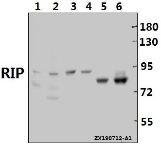 Western blot (WB) analysis of RIP pAb at 1:1000 dilution Lane1:C6 whole cell lysate(40ug) Lane2:3T3-L1 whole cell lysate(40ug) Lane3:The Brain tissue lysate of Mouse(40ug) Lane4:The Stomach tissue lysate of Rat(40ug) Lane5:DLD whole cell lysate(40ug) Lane6:SGC7901 whole cell lysate(40ug)
 -
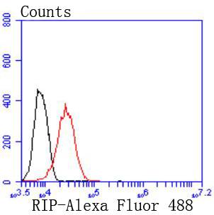 Flow cytometric analysis of 293 cells with RIP antibody at 1/50 dilution (red) compared with an unlabelled control (cells without incubation with primary antibody; black). Alexa Fluor 488-conjugated goat anti rabbit IgG was used as the secondary antibody
Bioworld Biotech only provide peptides for our antibodies and do not provide additional peptide customization services.
Price/Size :
USD 368/1mg/vial

Tips:
For phospho antibody, we provide phospho peptide(0.5mg) and non-phospho peptide(0.5mg).Describe :
Blocking peptides are peptides that bind specifically to the target antibody and block antibody binding. These peptide usually contains the epitope recognized by the antibody. Antibodies bound to the blocking peptide no longer bind to the epitope on the target protein. This mechanism is useful when non-specific binding is an issue, for example, in Western blotting (WB) and Immunohistochemistry (IHC). By comparing the staining from the blocked antibody versus the antibody alone, one can see which staining is specific; Specific binding will be absent from the western blot or IHC performed with the neutralized antibody.Formula:
Synthetic peptide was lyophilized with 100% acetonitrile and is supplied as a powder. Reconstitute with 0.1 ml DI water for a final concentration of 10 mg/ml.The purity is >90%,tested by HPLC and MS.
Storage:
The freeze-dried powder is more stable. For short time at 2-8°C. For long term storage store at -20°C.
Note :
This product is for research use only (RUO only). Not for use in diagnostic or therapeutic procedures.