
 p-VDR(S208) polyclonal antibody
  Datasheet
 COA
COA MSDS
MSDS SHIP
SHIP
Product Name :
p-VDR(S208) polyclonal antibody Background :
Although originally identified based on their roles in calcium and bone homeostasis, the vitamin D3 receptor (VDR/NR1I1) and its ligand 1-α, 25-dihydroxycholecalciferol [1α, 25(OH)2D3] are now recognized to exert biological effects in almost every tissue of the human body. Targets for vitamin D signaling include the central nervous system, skin, immune system, endocrine glands, kidney, and colon. At the cellular level, vitamin D signaling affects proliferation, differentiation, and apoptosis of both normal and transformed cells. Within the steroid receptor gene family, VDR belongs to the NR1I subfamily that also includes NR1I2/PXR and NR1I3/CAR. The human VDR gene is composed of 11 exons that encode six domains (A-F) of the full length VDR protein, which includes an N-terminal dual zinc finger DNA binding domain, a C-terminal ligand-binding activity domain, and an extensive unstructured region that links the two functional domains together . Upon 1α, 25(OH)2D3 binding to the hormone ligand-binding domain, VDR is stabilized by the phosphorylation of Ser51 in the DNA-binding domain by PKC, and Ser208 in the hinge region by casein kinase II. VDR associates with the retinoic acid receptor (RXR) through dimerization domains. The 1α, 25(OH)2D3-VDR-RXR complex binds to the vitamin D response elements (VDREs) in the promoters of target genes through the DNA-binding domain. Ligand-induced conformation changes in VDR results in the dissociation of the co-repressor, silencing-mediator for retinoid and thyroid hormone receptors (SMRT), and allows interaction of the VDR activation function (AF2) transactivation domain with transcriptional coactivators. Studies have shown that variable VDR expression is associated with different forms or stages of cancer and likely results from tissue-type variation in 1α, 25(OH)2D3 signaling. In the case of colon cancer, research indicates that VDR expression is relatively higher in hyperplastic colon polyps and during early tumorigenesis but diminishes in later stage, poorly differentiated tumors. Multiple studies suggest that 1α, 25(OH)2D3 may be an attractive target for development as a therapeutic anticancer agent. Product :
Rabbit IgG, 1mg/ml in PBS with 0.02% sodium azide, 50% glycerol, pH7.3. Storage&Stability :
Store at 4°C short term. Aliquot and store at -20°C long term. Avoid freeze-thaw cycles. Specificity :
p-VDR(S208) polyclonal antibody detects endogenous levels of p-VDR(S208) protein. Immunogen :
Synthetic peptide, corresponding to Human p-VDR(S208). Conjugate :
Unconjugated Modification :
Unmodification
-
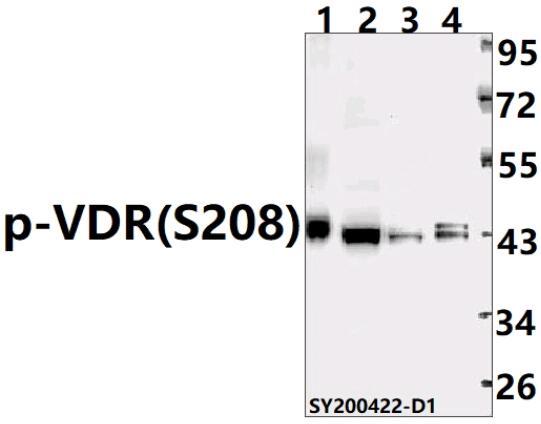 Western blot (WB) analysis of SUV39H2 polyclonal antibody at 1:500 dilution Lane1:SK-OVCAR3 whole cell lysate(40ug) Lane2:THP-1 whole cell lysate(40ug) Lane3:HEK293T whole cell lysate(40ug) Lane4:HCT116 whole cell lysate(40ug)
Bioworld Biotech only provide peptides for our antibodies and do not provide additional peptide customization services.
Price/Size :
USD 368/1mg/vial

Tips:
For phospho antibody, we provide phospho peptide(0.5mg) and non-phospho peptide(0.5mg).Describe :
Blocking peptides are peptides that bind specifically to the target antibody and block antibody binding. These peptide usually contains the epitope recognized by the antibody. Antibodies bound to the blocking peptide no longer bind to the epitope on the target protein. This mechanism is useful when non-specific binding is an issue, for example, in Western blotting (WB) and Immunohistochemistry (IHC). By comparing the staining from the blocked antibody versus the antibody alone, one can see which staining is specific; Specific binding will be absent from the western blot or IHC performed with the neutralized antibody.Formula:
Synthetic peptide was lyophilized with 100% acetonitrile and is supplied as a powder. Reconstitute with 0.1 ml DI water for a final concentration of 10 mg/ml.The purity is >90%,tested by HPLC and MS.
Storage:
The freeze-dried powder is more stable. For short time at 2-8°C. For long term storage store at -20°C.
Note :
This product is for research use only (RUO only). Not for use in diagnostic or therapeutic procedures.