
 Claudin 3 monoclonal antibody
  Datasheet
 COA
COA MSDS
MSDS SHIP
SHIP
Product Name :
Claudin 3 monoclonal antibody Background :
Tight junctions, or zonula occludens, form a continuous barrier to fluids across the epithelium and endothelium. They function in regulation of paracellular permeability and in the maintenance of cell polarity, blocking the movement of transmembrane proteins between the apical and the basolateral cell surfaces. Tight junctions are composed of claudin and occludin proteins, which join the junctions to the cytoskeleton . The claudin family is composed of 23 integral membrane proteins, and their expression, which varies among tissue types, may determine both the strength and properties of the epithelial barrier. Alteration in claudin protein expression pattern is associated with several types of cancer . Claudin-1 is expressed primarily in keratinocytes and normal mammary epithelial cells, but is absent or reduced in breast carcinomas and breast cancer cell lines . Claudin-3 is the product of an intronless gene (CLDN3) whose expression is markedly dysregulated in distinct cancer subtypes, including ovarian and breast carcinomas . Somewhat paradoxically, reduced claudin-3 expression is also a hallmark of a subset of breast tumors known as the claudin-low subtype, in which reduced claudin-3 expression was shown to correlate with a phenotype characteristic of cells undergoing epithelial-mesenchymal transition . Product :
Mouse IgG1. Liquid in PBS containing 50% glycerol, 0.2% BSA and 0.01% sodium azide. Storage&Stability :
Store at 4°C short term. Aliquot and store at -20°C long term. Avoid freeze-thaw cycles. Specificity :
Recognizes endogenous levels of Claudin 3 protein. Immunogen :
KLH-conjugated synthetic peptide encompassing a sequence within human Claudin 3. The exact sequence is proprietary. Conjugate :
Unconjugated Modification :
Unmodification
-

-
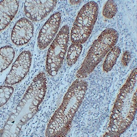 Immunohistochemical analysis of Claudin 3 staining in human colon formalin fixed paraffin embedded tissue section. The section was pre-treated using heat mediated antigen retrieval with sodium citrate buffer (pH 6.0). The section was then incubated with the antibody at room temperature and detected using an HRP conjugated compact polymer system. DAB was used as the chromogen. The section was then counterstained with haematoxylin and mounted with DPX.
 -
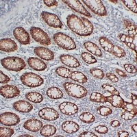 Immunohistochemical analysis of Claudin 3 staining in human colon formalin fixed paraffin embedded tissue section. The section was pre-treated using heat mediated antigen retrieval with sodium citrate buffer (pH 6.0). The section was then incubated with the antibody at room temperature and detected using an HRP conjugated compact polymer system. DAB was used as the chromogen. The section was then counterstained with haematoxylin and mounted with DPX.
Bioworld Biotech only provide peptides for our antibodies and do not provide additional peptide customization services.
Price/Size :
USD 368/1mg/vial

Tips:
For phospho antibody, we provide phospho peptide(0.5mg) and non-phospho peptide(0.5mg).Describe :
Blocking peptides are peptides that bind specifically to the target antibody and block antibody binding. These peptide usually contains the epitope recognized by the antibody. Antibodies bound to the blocking peptide no longer bind to the epitope on the target protein. This mechanism is useful when non-specific binding is an issue, for example, in Western blotting (WB) and Immunohistochemistry (IHC). By comparing the staining from the blocked antibody versus the antibody alone, one can see which staining is specific; Specific binding will be absent from the western blot or IHC performed with the neutralized antibody.Formula:
Synthetic peptide was lyophilized with 100% acetonitrile and is supplied as a powder. Reconstitute with 0.1 ml DI water for a final concentration of 10 mg/ml.The purity is >90%,tested by HPLC and MS.
Storage:
The freeze-dried powder is more stable. For short time at 2-8°C. For long term storage store at -20°C.
Note :
This product is for research use only (RUO only). Not for use in diagnostic or therapeutic procedures.