
 FGFR1 monoclonal antibody
  Datasheet
 COA
COA MSDS
MSDS SHIP
SHIP
Product Name :
FGFR1 monoclonal antibody Background :
Fibroblast growth factors (FGFs) produce mitogenic and angiogenic effects in target cells by signaling through cell surface receptor tyrosine kinases. There are four members of the FGF receptor family: FGFR1 (flg), FGFR2 (bek, KGFR), FGFR3, and FGFR4. Each receptor contains an extracellular ligand-binding domain, a transmembrane domain, and a cytoplasmic kinase domain . Following ligand binding and dimerization, the receptors are phosphorylated at specific tyrosine residues . Seven tyrosine residues in the cytoplasmic tail of FGFR1 can be phosphorylated: Tyr463, 583, 585, 653, 654, 730, and 766. Tyr653 and Tyr654 are important for catalytic activity of activated FGFR and are essential for signaling . The other phosphorylated tyrosine residues may provide docking sites for downstream signaling components, such as Crk and PLC. Product :
Mouse IgG1 kappa. Liquid in PBS, pH 7.3, 30% glycerol, and 0.01% sodium azide. Storage&Stability :
Store at 4°C short term. Aliquot and store at -20°C long term. Avoid freeze-thaw cycles. Specificity :
Recognizes endogenous levels of FGFR1 protein. Immunogen :
KLH-conjugated synthetic peptide encompassing a sequence within the C-term region of human FGFR1. The exact sequence is proprietary. Conjugate :
Unconjugated Modification :
Unmodification
-
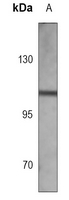 Western blot analysis of FGFR1 expression in Hela (A) whole cell lysates.
 -
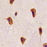 Immunohistochemical analysis of FGFR1 staining in human brain formalin fixed paraffin embedded tissue section. The section was pre-treated using heat mediated antigen retrieval with sodium citrate buffer (pH 6.0). The section was then incubated with the antibody at room temperature and detected using an HRP conjugated compact polymer system. DAB was used as the chromogen. The section was then counterstained with haematoxylin and mounted with DPX.
Bioworld Biotech only provide peptides for our antibodies and do not provide additional peptide customization services.
Price/Size :
USD 368/1mg/vial

Tips:
For phospho antibody, we provide phospho peptide(0.5mg) and non-phospho peptide(0.5mg).Describe :
Blocking peptides are peptides that bind specifically to the target antibody and block antibody binding. These peptide usually contains the epitope recognized by the antibody. Antibodies bound to the blocking peptide no longer bind to the epitope on the target protein. This mechanism is useful when non-specific binding is an issue, for example, in Western blotting (WB) and Immunohistochemistry (IHC). By comparing the staining from the blocked antibody versus the antibody alone, one can see which staining is specific; Specific binding will be absent from the western blot or IHC performed with the neutralized antibody.Formula:
Synthetic peptide was lyophilized with 100% acetonitrile and is supplied as a powder. Reconstitute with 0.1 ml DI water for a final concentration of 10 mg/ml.The purity is >90%,tested by HPLC and MS.
Storage:
The freeze-dried powder is more stable. For short time at 2-8°C. For long term storage store at -20°C.
Note :
This product is for research use only (RUO only). Not for use in diagnostic or therapeutic procedures.