
 RET monoclonal antibody
  Datasheet
 COA
COA MSDS
MSDS SHIP
SHIP
Product Name :
RET monoclonal antibody Background :
The Ret proto-oncogene is structurally related to the growing family of tyrosine kinase transmembrane receptors and is involved in GDNF signaling. By alternative splicing, two isoforms of the Ret proto-oncogene product are generated. The isoforms differ from each other by having either 9 or 51 carboxyterminal amino acids. The Ret gene products include two glycosylated proteins and, in Tunicamycin treated cells, a non-glycosylated protein consistent with the predicted Ret molecular weight based on sequence analysis. Tumorspecific rearrangements of the Ret proto-oncogene have been identified in papillary thyroid carcinomas leading to the formation of different transforming fusion proteins sharing the tyrosine kinase domain of Ret. In contrast to the Ret proto-oncogene, the rearranged forms are constitutively phosphorylated on tyrosine and are translocated from the membrane to the cytoplasm. Product :
Mouse IgM kappa. Supplied in crude ascites with 0.01% sodium azide. Storage&Stability :
Store at 4°C short term. Aliquot and store at -20°C long term. Avoid freeze-thaw cycles. Specificity :
Recognizes endogenous levels of RET protein. Immunogen :
Recombinant fusion protein of human RET. The exact sequence is proprietary. Conjugate :
Unconjugated Modification :
Unmodification
-
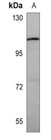 Western blot analysis of RET expression in A549 (A) whole cell lysates.
 -
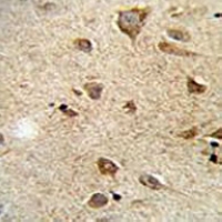 Immunohistochemical analysis of RET staining in human brain formalin fixed paraffin embedded tissue section. The section was pre-treated using heat mediated antigen retrieval with sodium citrate buffer (pH 6.0). The section was then incubated with the antibody at room temperature and detected using an HRP conjugated compact polymer system. DAB was used as the chromogen. The section was then counterstained with haematoxylin and mounted with DPX.
Bioworld Biotech only provide peptides for our antibodies and do not provide additional peptide customization services.
Price/Size :
USD 368/1mg/vial

Tips:
For phospho antibody, we provide phospho peptide(0.5mg) and non-phospho peptide(0.5mg).Describe :
Blocking peptides are peptides that bind specifically to the target antibody and block antibody binding. These peptide usually contains the epitope recognized by the antibody. Antibodies bound to the blocking peptide no longer bind to the epitope on the target protein. This mechanism is useful when non-specific binding is an issue, for example, in Western blotting (WB) and Immunohistochemistry (IHC). By comparing the staining from the blocked antibody versus the antibody alone, one can see which staining is specific; Specific binding will be absent from the western blot or IHC performed with the neutralized antibody.Formula:
Synthetic peptide was lyophilized with 100% acetonitrile and is supplied as a powder. Reconstitute with 0.1 ml DI water for a final concentration of 10 mg/ml.The purity is >90%,tested by HPLC and MS.
Storage:
The freeze-dried powder is more stable. For short time at 2-8°C. For long term storage store at -20°C.
Note :
This product is for research use only (RUO only). Not for use in diagnostic or therapeutic procedures.