
 PSMB8 monoclonal antibody
  Datasheet
 COA
COA MSDS
MSDS SHIP
SHIP
Product Name :
PSMB8 monoclonal antibody Background :
The 26S proteasome is a highly abundant proteolytic complex involved in the degradation of ubiquitinated substrate proteins. It consists largely of two sub-complexes, the 20S catalytic core particle (CP) and the 19S/PA700 regulatory particle (RP) that can cap either end of the CP. The CP consists of two stacked heteroheptameric β-rings (β1-7) that contain three catalytic β-subunits and are flanked on either side by two heteroheptameric α-rings (α1-7). The RP includes a base and a lid, each having multiple subunits. The base, in part, is composed of a heterohexameric ring of ATPase subunits belonging to the AAA (ATPases Associated with diverse cellular Activities) family. The ATPase subunits function to unfold the substrate and open the gate formed by the α-subunits, thus exposing the unfolded substrate to the catalytic β-subunits. The lid consists of ubiquitin receptors and DUBs that function in recruitment of ubiquitinated substrates and modification of ubiquitin chain topology. Other modulators of proteasome activity, such as PA28/11S REG, can also bind to the end of the 20S CP and activate it. Constitutively expressed core particle subunits PSMB5, PSMB7, and PSMB6 provide chymotrypsin-like, trypsin-like, and caspase-like activities, respectively. In immune cells involved in antigen presentation, these subunits are replaced by highly homologous, induced β-subunits to form the immunoproteasome. Proteasome subunit beta type-8 (PSMB8, LMP7) is expressed as a proenzyme that is cleaved to form the mature PSMB8 (LMP7) immunoproteasome core particle subunit. Interferon-γ induces expression of PSMB8, which functionally replaces the PSMB5 core particle subunit in immunoproteasome processing of MHC class I-restricted peptide antigens. Research studies suggest that reduced PSMB8 expression or expression of the non-functional LMP7-E1 isoform may impair immunoproteasome assembly, and that PSMB8 deficiency results in reduced MHC class I molecule expression. Inhibition of PSMB8 in murine rheumatoid arthritis models attenuates disease indicators, suggesting that PSMB8 is a potential therapeutic target in the treatment of some proinflammatory autoimmune diseases. Mutations in the corresponding PSMB8 gene can cause an autoinflammatory syndrome known as CANDLE Syndrome. Product :
Liquid in PBS containing 50% glycerol, 0.5% BSA and 0.02% sodium azide, pH 7.3. Storage&Stability :
Store at 4°C short term. Aliquot and store at -20°C long term. Avoid freeze-thaw cycles. Specificity :
Recognizes endogenous levels of PSMB8 protein. Immunogen :
Purified recombinant fragment of human PSMB8 expressed in E. Coli. Conjugate :
Unconjugated Modification :
Unmodification
-
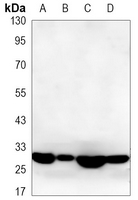 Western blot analysis of PSMB8 expression in C6 (A), Hela (B), Molt4 (C), Raji (D) whole cell lysates.
 -
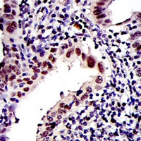 Immunohistochemical analysis of PSMB8 staining in human intima cancer formalin fixed paraffin embedded tissue section. The section was pre-treated using heat mediated antigen retrieval with sodium citrate buffer (pH 6.70). The section was then incubated with the antibody at room temperature and detected using an HRP conjugated compact polymer system. DAB was used as the chromogen. The section was then counterstained with haematoxylin and mounted with DPX.
Bioworld Biotech only provide peptides for our antibodies and do not provide additional peptide customization services.
Price/Size :
USD 368/1mg/vial

Tips:
For phospho antibody, we provide phospho peptide(0.5mg) and non-phospho peptide(0.5mg).Describe :
Blocking peptides are peptides that bind specifically to the target antibody and block antibody binding. These peptide usually contains the epitope recognized by the antibody. Antibodies bound to the blocking peptide no longer bind to the epitope on the target protein. This mechanism is useful when non-specific binding is an issue, for example, in Western blotting (WB) and Immunohistochemistry (IHC). By comparing the staining from the blocked antibody versus the antibody alone, one can see which staining is specific; Specific binding will be absent from the western blot or IHC performed with the neutralized antibody.Formula:
Synthetic peptide was lyophilized with 100% acetonitrile and is supplied as a powder. Reconstitute with 0.1 ml DI water for a final concentration of 10 mg/ml.The purity is >90%,tested by HPLC and MS.
Storage:
The freeze-dried powder is more stable. For short time at 2-8°C. For long term storage store at -20°C.
Note :
This product is for research use only (RUO only). Not for use in diagnostic or therapeutic procedures.