
 PAK2 monoclonal antibody
  Datasheet
 COA
COA MSDS
MSDS SHIP
SHIP
Product Name :
PAK2 monoclonal antibody Background :
The p21-activated kinase (PAK) family of serine/threonine kinases is engaged in multiple cellular processes, including cytoskeletal reorganization, MAPK signaling, apoptotic signaling, control of phagocyte NADPH oxidase, and growth factor-induced neurite outgrowth. Several mechanisms that induce PAK activity have been reported. Binding of Rac/Cdc42 to the CRIB (or PBD) domain near the amino terminus of PAK causes autophosphorylation and conformational changes in PAK. Phosphorylation of PAK1 at Thr423 by PDK induces activation of PAK1. Several autophosphorylation sites have been identified, including Ser199 and Ser204 of PAK1, and Ser192 and Ser197 of PAK2. Because the autophosphorylation sites are located in the amino-terminal inhibitory domain, it has been hypothesized that modification in this region prevents the kinase from reverting to an inactive conformation. Research indicates that phosphorylation at Ser144 of PAK1 or Ser139 of PAK3 (located in the kinase inhibitory domain) affects kinase activity. Phosphorylation at Ser21 of PAK1 or Ser20 of PAK2 regulates binding with the adaptor protein Nck. PAK4, PAK5/7, and PAK6 have lower sequence similarity with PAK1-3 in the amino-terminal regulatory region. Phosphorylation at Ser474 of PAK4, a site analogous to Thr423 of PAK1, may play a pivotal role in regulating the activity and function of PAK4. PAK family members are widely expressed, and often overexpressed in human cancer. Product :
Liquid in PBS containing 50% glycerol, 0.5% BSA and 0.02% sodium azide, pH 7.3. Storage&Stability :
Store at 4°C short term. Aliquot and store at -20°C long term. Avoid freeze-thaw cycles. Specificity :
Recognizes endogenous levels of PAK2 protein. Immunogen :
Purified recombinant fragment of PAK2 expressed in E. Coli. Conjugate :
Unconjugated Modification :
Unmodification
-
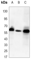 Western blot analysis of PAK2 expression in Jurkat (A), MCF7 (B), Hela (C) whole cell lysates.
 -
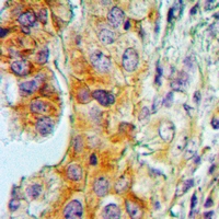 Immunohistochemical analysis of PAK2 staining in human lung cancer formalin fixed paraffin embedded tissue section. The section was pre-treated using heat mediated antigen retrieval with sodium citrate buffer (pH 6.102). The section was then incubated with the antibody at room temperature and detected using an HRP conjugated compact polymer system. DAB was used as the chromogen. The section was then counterstained with haematoxylin and mounted with DPX.
Bioworld Biotech only provide peptides for our antibodies and do not provide additional peptide customization services.
Price/Size :
USD 368/1mg/vial

Tips:
For phospho antibody, we provide phospho peptide(0.5mg) and non-phospho peptide(0.5mg).Describe :
Blocking peptides are peptides that bind specifically to the target antibody and block antibody binding. These peptide usually contains the epitope recognized by the antibody. Antibodies bound to the blocking peptide no longer bind to the epitope on the target protein. This mechanism is useful when non-specific binding is an issue, for example, in Western blotting (WB) and Immunohistochemistry (IHC). By comparing the staining from the blocked antibody versus the antibody alone, one can see which staining is specific; Specific binding will be absent from the western blot or IHC performed with the neutralized antibody.Formula:
Synthetic peptide was lyophilized with 100% acetonitrile and is supplied as a powder. Reconstitute with 0.1 ml DI water for a final concentration of 10 mg/ml.The purity is >90%,tested by HPLC and MS.
Storage:
The freeze-dried powder is more stable. For short time at 2-8°C. For long term storage store at -20°C.
Note :
This product is for research use only (RUO only). Not for use in diagnostic or therapeutic procedures.