
 FUBP1 Rabbit monoclonal antibody
  Datasheet
 COA
COA MSDS
MSDS SHIP
SHIP
Product Name :
FUBP1 Rabbit monoclonal antibody Background :
The protein encoded by this gene is a single stranded DNA-binding protein that binds to multiple DNA elements, including the far upstream element (FUSE) located upstream of cmyc. Binding to FUSE occurs on the non-coding strand, and is important to the regulation of c-myc in undifferentiated cells. This protein contains three domains, an amphipathic helix Nterminal domain, a DNA-binding central domain, and a C-terminal transactivation domain that contains three tyrosine-rich motifs. The N-terminal domain is thought to repress the activity of the C-terminal domain. This protein is also thought to bind RNA, and contains 3'-5' helicase activity with in vitro activity on both DNA-DNA and RNA-RNA duplexes. Aberrant expression of this gene has been found in malignant tissues, and this gene is important to neural system and lung development. Binding of this protein to viral RNA is thought to play a role in several viral diseases, including hepatitis C and hand, foot and mouth disease. Alternative splicing results in multiple transcript variants. Product :
Liquid in 50mM Tris-Glycine (pH 7.4), 0.15M NaCl, 50% Glycerol, 0.01% Sodium azide and 0.05% BSA. Storage&Stability :
Store at 4°C short term. Aliquot and store at -20°C long term. Avoid freeze-thaw cycles. Specificity :
Recognizes endogenous levels of FUBP1 protein. Immunogen :
A synthetic peptide of human FUBP1 Conjugate :
Unconjugated Modification :
Unmodification
-
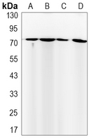 Western blot analysis of FUBP1 expression in mouse spleen (A), K562 (B), rat brain (C), Hela (D) whole cell lysates.
 -
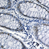 Immunohistochemical analysis of FUBP1 staining in human colon cancer formalin fixed paraffin embedded tissue section. The section was pre-treated using heat mediated antigen retrieval with sodium citrate buffer (pH 6.57). The section was then incubated with the antibody at room temperature and detected using an HRP conjugated compact polymer system. DAB was used as the chromogen. The section was then counterstained with haematoxylin and mounted with DPX.
Bioworld Biotech only provide peptides for our antibodies and do not provide additional peptide customization services.
Price/Size :
USD 368/1mg/vial

Tips:
For phospho antibody, we provide phospho peptide(0.5mg) and non-phospho peptide(0.5mg).Describe :
Blocking peptides are peptides that bind specifically to the target antibody and block antibody binding. These peptide usually contains the epitope recognized by the antibody. Antibodies bound to the blocking peptide no longer bind to the epitope on the target protein. This mechanism is useful when non-specific binding is an issue, for example, in Western blotting (WB) and Immunohistochemistry (IHC). By comparing the staining from the blocked antibody versus the antibody alone, one can see which staining is specific; Specific binding will be absent from the western blot or IHC performed with the neutralized antibody.Formula:
Synthetic peptide was lyophilized with 100% acetonitrile and is supplied as a powder. Reconstitute with 0.1 ml DI water for a final concentration of 10 mg/ml.The purity is >90%,tested by HPLC and MS.
Storage:
The freeze-dried powder is more stable. For short time at 2-8°C. For long term storage store at -20°C.
Note :
This product is for research use only (RUO only). Not for use in diagnostic or therapeutic procedures.