
 SMAD3 Rabbit monoclonal antibody
  Datasheet
 COA
COA MSDS
MSDS SHIP
SHIP
Product Name :
SMAD3 Rabbit monoclonal antibody Background :
Members of the Smad family of signal transduction molecules are components of a critical intracellular pathway that transmit TGF-β signals from the cell surface into the nucleus. Three distinct classes of Smads have been defined: the receptor-regulated Smads (R-Smads), which include Smad1, 2, 3, 5, and 8; the common-mediator Smad (co-Smad), Smad4; and the antagonistic or inhibitory Smads (I-Smads), Smad6 and 7. Activated type I receptors associate with specific R-Smads and phosphorylate them on a conserved carboxy-terminal SSXS motif. The phosphorylated R-Smad dissociates from the receptor and forms a heteromeric complex with the co-Smad (Smad4), allowing translocation of the complex to the nucleus. Once in the nucleus, Smads can target a variety of DNA binding proteins to regulate transcriptional responses. Following stimulation by TGF-β, Smad2 and Smad3 become phosphorylated at their carboxyl termini (Ser465 and 467 on Smad2; Ser423 and 425 on Smad3) by TGF-β Receptor I. Phosphorylated Smad 2/3 can complex with Smad4, translocate to the nucleus and regulate gene expression. Product :
Liquid in 50mM Tris-Glycine (pH 7.4), 0.15M NaCl, 50% Glycerol, 0.01% Sodium azide and 0.05% BSA. Storage&Stability :
Store at 4°C short term. Aliquot and store at -20°C long term. Avoid freeze-thaw cycles. Specificity :
Recognizes endogenous levels of SMAD3 protein. Immunogen :
A synthetic peptide of human Smad3 Conjugate :
Unconjugated Modification :
Unmodification
-
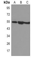 Western blot analysis of SMAD3 expression in Jurkat (A), C6 (B), Hela (C) whole cell lysates.
 -
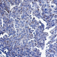 Immunohistochemical analysis of SMAD3 staining in human breast carcinoma formalin fixed paraffin embedded tissue section. The section was pre-treated using heat mediated antigen retrieval with sodium citrate buffer (pH 6.5). The section was then incubated with the antibody at room temperature and detected using an HRP conjugated compact polymer system. DAB was used as the chromogen. The section was then counterstained with haematoxylin and mounted with DPX.
Bioworld Biotech only provide peptides for our antibodies and do not provide additional peptide customization services.
Price/Size :
USD 368/1mg/vial

Tips:
For phospho antibody, we provide phospho peptide(0.5mg) and non-phospho peptide(0.5mg).Describe :
Blocking peptides are peptides that bind specifically to the target antibody and block antibody binding. These peptide usually contains the epitope recognized by the antibody. Antibodies bound to the blocking peptide no longer bind to the epitope on the target protein. This mechanism is useful when non-specific binding is an issue, for example, in Western blotting (WB) and Immunohistochemistry (IHC). By comparing the staining from the blocked antibody versus the antibody alone, one can see which staining is specific; Specific binding will be absent from the western blot or IHC performed with the neutralized antibody.Formula:
Synthetic peptide was lyophilized with 100% acetonitrile and is supplied as a powder. Reconstitute with 0.1 ml DI water for a final concentration of 10 mg/ml.The purity is >90%,tested by HPLC and MS.
Storage:
The freeze-dried powder is more stable. For short time at 2-8°C. For long term storage store at -20°C.
Note :
This product is for research use only (RUO only). Not for use in diagnostic or therapeutic procedures.