
 HMGB1 Rabbit monoclonal antibody
  Datasheet
 COA
COA MSDS
MSDS SHIP
SHIP
Product Name :
HMGB1 Rabbit monoclonal antibody Background :
High mobility group protein B1 (HMGB1) belongs to a family of highly conserved proteins that contain HMG box domains. All three family members (HMGB1, HMGB2, and HMGB3) contain two HMG box domains and a C-terminal acidic domain. HMGB1 is a widely expressed and highly abundant protein. HMGB2 is widely expressed during embryonic development, but is restricted to lymphoid organs and testis in adult animals. HMGB3 is only expressed during embryogenesis. While expression varies, the biochemical properties of the different family members may be indistinguishable. The HMG box domains facilitate the binding of HMGB proteins to the minor groove of DNA, which results in local bending of the DNA double helix. HMGB proteins are recruited by and help facilitate the assembly of site-specific DNA binding proteins to their cognate binding sites in chromatin. For example, HMGB1 facilitates the binding of Hox proteins, Oct-1, p53, Rel proteins, and steroid hormone receptor proteins to their target gene promoters. In addition to their functions in the nucleus, HMGB proteins play a significant role in extracellular signaling associated with inflammation. HMGB1 is massively released into the extracellular environment during cell necrosis, but not apoptosis. Extracellular HMGB1 "alarms" the innate immune system by acting as a chemoattractant for inflammatory leukocytes, smooth muscle cells, and stem cells, functioning as an immune adjuvant for soluble and particulate antigens, and triggering activation of T cells and dendritic cells. In addition, activated monocytes, macrophages and, dendritic cells also secrete HMGB1, forming a positive feedback loop that results in the release of additional cytokines and neutrophils. Hypoxia has also been shown to cause the release of HMGB1 in the liver, and some studies suggest a role for extracellular HMGB1 in tumor homeostasis. Product :
Liquid in 50mM Tris-Glycine (pH 7.4), 0.15M NaCl, 50% Glycerol, 0.01% Sodium azide and 0.05% BSA. Storage&Stability :
Store at 4°C short term. Aliquot and store at -20°C long term. Avoid freeze-thaw cycles. Specificity :
Recognizes endogenous levels of HMGB1 protein. Immunogen :
A synthetic peptide of human HMGB1 Conjugate :
Unconjugated Modification :
Unmodification
-
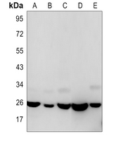 Western blot analysis of HMGB1 expression in K562 (A), rat brain (B), C6 (C), NIH3T3 (D), Hela (E) whole cell lysates.
 -
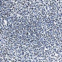 Immunohistochemical analysis of HMGB1 staining in human tonsil formalin fixed paraffin embedded tissue section. The section was pre-treated using heat mediated antigen retrieval with sodium citrate buffer (pH 6.111). The section was then incubated with the antibody at room temperature and detected using an HRP conjugated compact polymer system. DAB was used as the chromogen. The section was then counterstained with haematoxylin and mounted with DPX.
Bioworld Biotech only provide peptides for our antibodies and do not provide additional peptide customization services.
Price/Size :
USD 368/1mg/vial

Tips:
For phospho antibody, we provide phospho peptide(0.5mg) and non-phospho peptide(0.5mg).Describe :
Blocking peptides are peptides that bind specifically to the target antibody and block antibody binding. These peptide usually contains the epitope recognized by the antibody. Antibodies bound to the blocking peptide no longer bind to the epitope on the target protein. This mechanism is useful when non-specific binding is an issue, for example, in Western blotting (WB) and Immunohistochemistry (IHC). By comparing the staining from the blocked antibody versus the antibody alone, one can see which staining is specific; Specific binding will be absent from the western blot or IHC performed with the neutralized antibody.Formula:
Synthetic peptide was lyophilized with 100% acetonitrile and is supplied as a powder. Reconstitute with 0.1 ml DI water for a final concentration of 10 mg/ml.The purity is >90%,tested by HPLC and MS.
Storage:
The freeze-dried powder is more stable. For short time at 2-8°C. For long term storage store at -20°C.
Note :
This product is for research use only (RUO only). Not for use in diagnostic or therapeutic procedures.