
 E Cadherin Rabbit monoclonal antibody
  Datasheet
 COA
COA MSDS
MSDS SHIP
SHIP
Product Name :
E Cadherin Rabbit monoclonal antibody Background :
Cadherins are a superfamily of transmembrane glycoproteins that contain cadherin repeats of approximately 100 residues in their extracellular domain. Cadherins mediate calcium-dependent cell-cell adhesion and play critical roles in normal tissue development. The classic cadherin subfamily includes N-, P-, R-, B-, and E-cadherins, as well as about ten other members that are found in adherens junctions, a cellular structure near the apical surface of polarized epithelial cells. The cytoplasmic domain of classical cadherins interacts with β-catenin, γ-catenin (also called plakoglobin), and p120 catenin. β-catenin and γ-catenin associate with α-catenin, which links the cadherin-catenin complex to the actin cytoskeleton. While β- and γ-catenin play structural roles in the junctional complex, p120 regulates cadherin adhesive activity and trafficking. Investigators consider E-cadherin an active suppressor of invasion and growth of many epithelial cancers. Research studies indicate that cancer cells have upregulated N-cadherin in addition to loss of E-cadherin. This change in cadherin expression is called the "cadherin switch." N-cadherin cooperates with the FGF receptor, leading to overexpression of MMP-9 and cellular invasion. Research studies have shown that in endothelial cells, VE-cadherin signaling, expression, and localization correlate with vascular permeability and tumor angiogenesis. Investigators have also demonstrated that expression of P-cadherin, which is normally present in epithelial cells, is also altered in ovarian and other human cancers. Product :
Liquid in 50mM Tris-Glycine (pH 7.4), 0.15M NaCl, 50% Glycerol, 0.01% Sodium azide and 0.05% BSA. Storage&Stability :
Store at 4°C short term. Aliquot and store at -20°C long term. Avoid freeze-thaw cycles. Specificity :
Recognizes endogenous levels of E Cadherin protein. Immunogen :
A synthetic peptide of human E Cadherin Conjugate :
Unconjugated Modification :
Unmodification
-
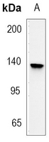 Western blot analysis of E Cadherin expression in PC3 (A) whole cell lysates.
 -
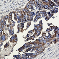 Immunohistochemical analysis of E Cadherin staining in human cholangiocarcinoma formalin fixed paraffin embedded tissue section. The section was pre-treated using heat mediated antigen retrieval with sodium citrate buffer (pH 6.36). The section was then incubated with the antibody at room temperature and detected using an HRP conjugated compact polymer system. DAB was used as the chromogen. The section was then counterstained with haematoxylin and mounted with DPX.
Bioworld Biotech only provide peptides for our antibodies and do not provide additional peptide customization services.
Price/Size :
USD 368/1mg/vial

Tips:
For phospho antibody, we provide phospho peptide(0.5mg) and non-phospho peptide(0.5mg).Describe :
Blocking peptides are peptides that bind specifically to the target antibody and block antibody binding. These peptide usually contains the epitope recognized by the antibody. Antibodies bound to the blocking peptide no longer bind to the epitope on the target protein. This mechanism is useful when non-specific binding is an issue, for example, in Western blotting (WB) and Immunohistochemistry (IHC). By comparing the staining from the blocked antibody versus the antibody alone, one can see which staining is specific; Specific binding will be absent from the western blot or IHC performed with the neutralized antibody.Formula:
Synthetic peptide was lyophilized with 100% acetonitrile and is supplied as a powder. Reconstitute with 0.1 ml DI water for a final concentration of 10 mg/ml.The purity is >90%,tested by HPLC and MS.
Storage:
The freeze-dried powder is more stable. For short time at 2-8°C. For long term storage store at -20°C.
Note :
This product is for research use only (RUO only). Not for use in diagnostic or therapeutic procedures.