
 YAP1 polyclonal antibody
  Datasheet
 COA
COA MSDS
MSDS SHIP
SHIP
Product Name :
YAP1 polyclonal antibody Background :
YAP (Yes-associated protein, YAP65) was first identified based on its ability to associate with the SH3 domain of Yes. It also binds to other SH3 domain-containing proteins such as Nck, Crk, Src, and Abl. In addition to the SH3 binding motif, YAP contains a PDZ interaction motif, a coiled-coil domain, and WW domains. While initial studies of YAP all pointed towards a role in anchoring and targeting to specific subcellular compartments, subsequent studies showed that YAP is a transcriptional co-activator by virtue of its WW domain interacting with the PY motif (PPxY) of the transcription factor PEBP2 and other transcription factors. In its capacity as a transcriptional co-activator, YAP is now widely recognized as a central mediator of the Hippo Pathway, which plays a fundamental and widely conserved role in regulating tissue growth and organ size. Phosphorylation at multiple sites (e.g., Ser109, Ser127) by LATS kinases promotes YAP translocation from the nucleus to the cytoplasm, where it is sequestered through association with 14-3-3 proteins. These LATS-driven phosphorylation events serve to prime YAP for subsequent phosphorylation by CK1δ/ε in an adjacent phosphodegron, triggering proteasomal degradation of YAP. Product :
Liquid in 0.42% Potassium phosphate, 0.87% Sodium chloride, pH 7.3, 30% glycerol, and 0.01% sodium azide. Storage&Stability :
Store at 4°C short term. Aliquot and store at -20°C long term. Avoid freeze-thaw cycles. Specificity :
Recognizes endogenous levels of YAP1 protein. Immunogen :
Recombinant protein corresponding to human YAP1. The exact sequence is proprietary. Conjugate :
Unconjugated Modification :
Unmodification
-
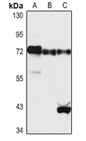 Western blot analysis of YAP1 expression in HEK293T (A), NIH3T3 (B), rat liver (C) whole cell lysates.
 -
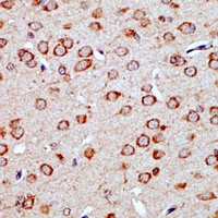 Immunohistochemical analysis of YAP1 staining in rat brain formalin fixed paraffin embedded tissue section. The section was pre-treated using heat mediated antigen retrieval with sodium citrate buffer (pH 6.0). The section was then incubated with the antibody at room temperature and detected using an HRP conjugated compact polymer system. DAB was used as the chromogen. The section was then counterstained with haematoxylin and mounted with DPX.
Bioworld Biotech only provide peptides for our antibodies and do not provide additional peptide customization services.
Price/Size :
USD 368/1mg/vial

Tips:
For phospho antibody, we provide phospho peptide(0.5mg) and non-phospho peptide(0.5mg).Describe :
Blocking peptides are peptides that bind specifically to the target antibody and block antibody binding. These peptide usually contains the epitope recognized by the antibody. Antibodies bound to the blocking peptide no longer bind to the epitope on the target protein. This mechanism is useful when non-specific binding is an issue, for example, in Western blotting (WB) and Immunohistochemistry (IHC). By comparing the staining from the blocked antibody versus the antibody alone, one can see which staining is specific; Specific binding will be absent from the western blot or IHC performed with the neutralized antibody.Formula:
Synthetic peptide was lyophilized with 100% acetonitrile and is supplied as a powder. Reconstitute with 0.1 ml DI water for a final concentration of 10 mg/ml.The purity is >90%,tested by HPLC and MS.
Storage:
The freeze-dried powder is more stable. For short time at 2-8°C. For long term storage store at -20°C.
Note :
This product is for research use only (RUO only). Not for use in diagnostic or therapeutic procedures.