
 AMPD1 polyclonal antibody
  Datasheet
 COA
COA MSDS
MSDS SHIP
SHIP
Product Name :
AMPD1 polyclonal antibody Background :
Adenosine monophosphate deaminase 1 catalyzes the deamination of AMP to IMP in skeletal muscle and plays an important role in the purine nucleotide cycle. Two other genes have been identified, AMPD2 and AMPD3, for the liver- and erythocyte-specific isoforms, respectively. Deficiency of the muscle-specific enzyme is apparently a common cause of exercise-induced myopathy and probably the most common cause of metabolic myopathy in the human. Alternatively spliced transcript variants encoding different isoforms have been identified in this gene. Product :
1mg/ml in PBS with 0.02% sodium azide, 50% glycerol, pH7.2 Storage&Stability :
Store at 4°C short term. Aliquot and store at -20°C long term. Avoid freeze-thaw cycles. Specificity :
Unmodification Immunogen :
Recombinant fusion protein of human AMPD1(NP_001166097.1). Conjugate :
Unconjugated Modification :
Unmodification
-
 Western blot analysis of extracts of various cell lines, using AMPD1 antibody at 1:3000 dilution.
Secondary antibody: HRP Goat Anti-Rabbit IgG at 1:10000 dilution.
Lysates/proteins: 25ug per lane.
Blocking buffer: 3% nonfat dry milk in TBST.
Detection: ECL Basic Kit .
Exposure time: 90s. -
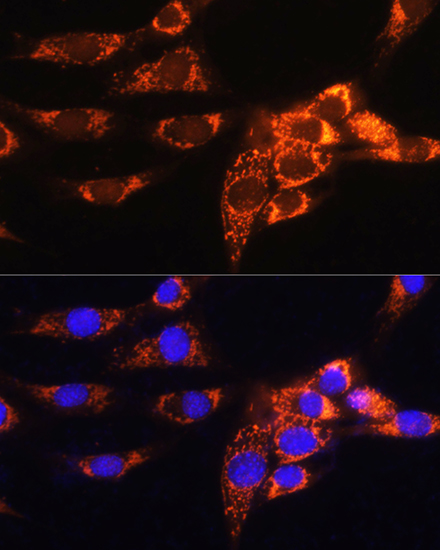 Immunofluorescence analysis of NIH/3T3 cells using AMPD1 Rabbit pAb at dilution of 1:100. Blue: DAPI for nuclear staining.
 -
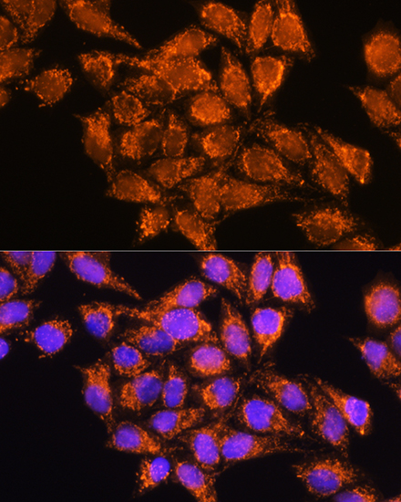 Immunofluorescence analysis of NIH/3T3 cells using AMPD1 Rabbit pAb at dilution of 1:100. Blue: DAPI for nuclear staining.
 -
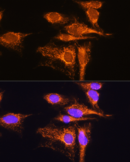 Immunofluorescence analysis of NIH/3T3 cells using AMPD1 Rabbit pAb at dilution of 1:100. Blue: DAPI for nuclear staining.
Bioworld Biotech only provide peptides for our antibodies and do not provide additional peptide customization services.
Price/Size :
USD 368/1mg/vial

Tips:
For phospho antibody, we provide phospho peptide(0.5mg) and non-phospho peptide(0.5mg).Describe :
Blocking peptides are peptides that bind specifically to the target antibody and block antibody binding. These peptide usually contains the epitope recognized by the antibody. Antibodies bound to the blocking peptide no longer bind to the epitope on the target protein. This mechanism is useful when non-specific binding is an issue, for example, in Western blotting (WB) and Immunohistochemistry (IHC). By comparing the staining from the blocked antibody versus the antibody alone, one can see which staining is specific; Specific binding will be absent from the western blot or IHC performed with the neutralized antibody.Formula:
Synthetic peptide was lyophilized with 100% acetonitrile and is supplied as a powder. Reconstitute with 0.1 ml DI water for a final concentration of 10 mg/ml.The purity is >90%,tested by HPLC and MS.
Storage:
The freeze-dried powder is more stable. For short time at 2-8°C. For long term storage store at -20°C.
Note :
This product is for research use only (RUO only). Not for use in diagnostic or therapeutic procedures.