
 UFL1 polyclonal antibody
  Datasheet
 COA
COA MSDS
MSDS SHIP
SHIP
Product Name :
UFL1 polyclonal antibody Background :
E3 protein ligase that mediates ufmylation, the covalent attachment of the ubiquitin-like modifier UFM1 to lysine residues on target proteins, and which plays a key role in reticulophagy (also called ER-phagy induced in response to endoplasmic reticulum stress. In response to endoplasmic reticulum stress, recruited to the endoplasmic reticulum membrane by DDRGK1, and mediates ufmylation of proteins such as RPN1 and RPL26/uL24, thereby promoting reticulophagy of endoplasmic reticulum sheets. Ufmylation-dependent reticulophagy inhibits the unfolded protein response (UPR via ERN1/IRE1-alpha. Ufmylation in response to endoplasmic reticulum stress is essential for processes such as hematopoiesis, blood vessel morphogenesis or inflammatory response. Regulates inflammation in response to endoplasmic reticulum stress by promoting reticulophagy, leading to inhibit the activity of the NF-kappa-B transcription factor (By similarity. Product :
1mg/ml in PBS with 0.02% sodium azide, 50% glycerol, pH7.2 Storage&Stability :
Store at 4°C short term. Aliquot and store at -20°C long term. Avoid freeze-thaw cycles. Specificity :
Unmodification Immunogen :
Recombinant fusion protein of human UFL1(NP_056138.1). Conjugate :
Unconjugated Modification :
Unmodification
-
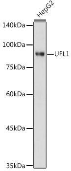 Western blot analysis of extracts of HepG2 cells, using UFL1 antibody at 1:1000 dilution.
Secondary antibody: HRP Goat Anti-Rabbit IgG at 1:10000 dilution.
Lysates/proteins: 25ug per lane.
Blocking buffer: 3% nonfat dry milk in TBST.
Detection: ECL Basic Kit .
Exposure time: 1s. -
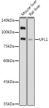 Western blot analysis of extracts of various cell lines, using UFL1 antibody at 1:1000 dilution.
Secondary antibody: HRP Goat Anti-Rabbit IgG at 1:10000 dilution.
Lysates/proteins: 25ug per lane.
Blocking buffer: 3% nonfat dry milk in TBST.
Detection: ECL Basic Kit .
Exposure time: 3s. -
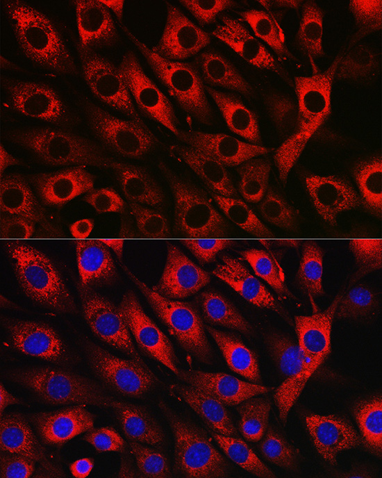 Western blot analysis of extracts of various cell lines, using UFL1 antibody at 1:1000 dilution.
Secondary antibody: HRP Goat Anti-Rabbit IgG at 1:10000 dilution.
Lysates/proteins: 25ug per lane.
Blocking buffer: 3% nonfat dry milk in TBST.
Detection: ECL Basic Kit .
Exposure time: 3s. -
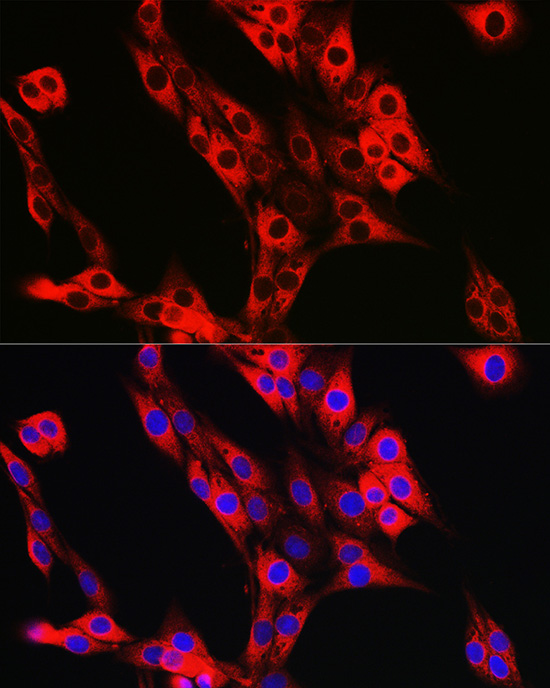 Western blot analysis of extracts of various cell lines, using UFL1 antibody at 1:1000 dilution.
Secondary antibody: HRP Goat Anti-Rabbit IgG at 1:10000 dilution.
Lysates/proteins: 25ug per lane.
Blocking buffer: 3% nonfat dry milk in TBST.
Detection: ECL Basic Kit .
Exposure time: 3s.
Bioworld Biotech only provide peptides for our antibodies and do not provide additional peptide customization services.
Price/Size :
USD 368/1mg/vial

Tips:
For phospho antibody, we provide phospho peptide(0.5mg) and non-phospho peptide(0.5mg).Describe :
Blocking peptides are peptides that bind specifically to the target antibody and block antibody binding. These peptide usually contains the epitope recognized by the antibody. Antibodies bound to the blocking peptide no longer bind to the epitope on the target protein. This mechanism is useful when non-specific binding is an issue, for example, in Western blotting (WB) and Immunohistochemistry (IHC). By comparing the staining from the blocked antibody versus the antibody alone, one can see which staining is specific; Specific binding will be absent from the western blot or IHC performed with the neutralized antibody.Formula:
Synthetic peptide was lyophilized with 100% acetonitrile and is supplied as a powder. Reconstitute with 0.1 ml DI water for a final concentration of 10 mg/ml.The purity is >90%,tested by HPLC and MS.
Storage:
The freeze-dried powder is more stable. For short time at 2-8°C. For long term storage store at -20°C.
Note :
This product is for research use only (RUO only). Not for use in diagnostic or therapeutic procedures.