
 SLC4A1 polyclonal antibody
  Datasheet
 COA
COA MSDS
MSDS SHIP
SHIP
Product Name :
SLC4A1 polyclonal antibody Background :
The protein encoded by this gene is part of the anion exchanger (AE) family and is expressed in the erythrocyte plasma membrane, where it functions as a chloride/bicarbonate exchanger involved in carbon dioxide transport from tissues to lungs. The protein comprises two domains that are structurally and functionally distinct. The N-terminal 40kDa domain is located in the cytoplasm and acts as an attachment site for the red cell skeleton by binding ankyrin. The glycosylated C-terminal membrane-associated domain contains 12-14 membrane spanning segments and carries out the stilbene disulphonate-sensitive exchange transport of anions. The cytoplasmic tail at the extreme C-terminus of the membrane domain binds carbonic anhydrase II. The encoded protein associates with the red cell membrane protein glycophorin A and this association promotes the correct folding and translocation of the exchanger. This protein is predominantly dimeric but forms tetramers in the presence of ankyrin. Many mutations in this gene are known in man, and these mutations can lead to two types of disease: destabilization of red cell membrane leading to hereditary spherocytosis, and defective kidney acid secretion leading to distal renal tubular acidosis. Other mutations that do not give rise to disease result in novel blood group antigens, which form the Diego blood group system.One null mutation in this gene is known, resulting in very severe anemia and nephrocalcinosis. Product :
1mg/ml in PBS with 0.02% sodium azide, 50% glycerol, pH7.2 Storage&Stability :
Store at 4°C short term. Aliquot and store at -20°C long term. Avoid freeze-thaw cycles. Specificity :
Unmodification Immunogen :
Recombinant fusion protein of human SLC4A1(NP_000333.1). Conjugate :
Unconjugated Modification :
Unmodification
-
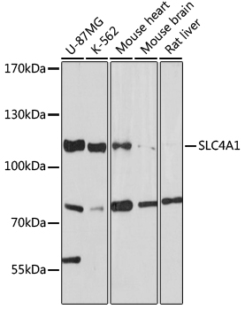 Western blot analysis of extracts of various cell lines, using SLC4A1 antibody at 1:1000 dilution.
Secondary antibody: HRP Goat Anti-Rabbit IgG at 1:10000 dilution.
Lysates/proteins: 25ug per lane.
Blocking buffer: 3% nonfat dry milk in TBST.
Detection: ECL Basic Kit .
Exposure time: 30s. -
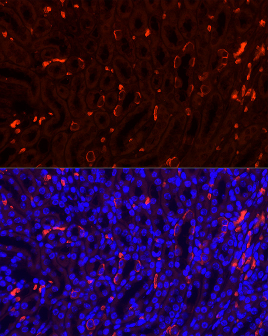 Immunofluorescence analysis of Mouse kidney using SLC4A1 antibody at dilution of 1:100. Blue: DAPI for nuclear staining.
 -
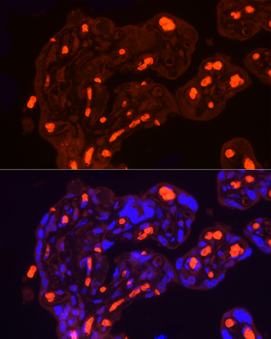 Immunofluorescence analysis of Mouse kidney using SLC4A1 antibody at dilution of 1:100. Blue: DAPI for nuclear staining.
 -
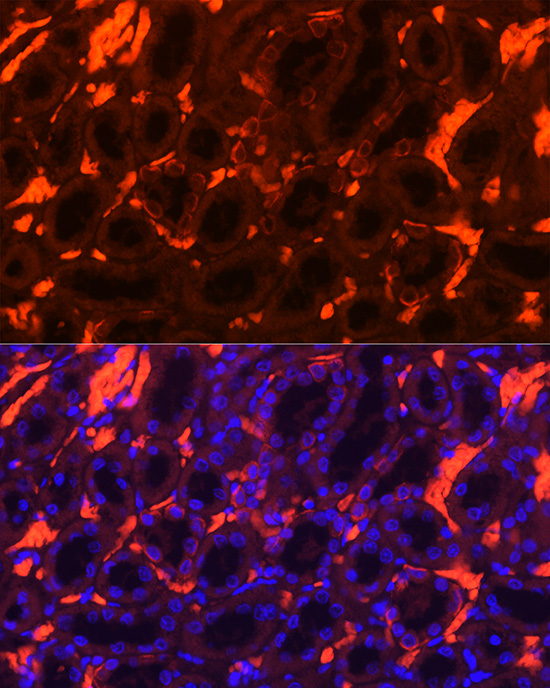 Immunofluorescence analysis of Mouse kidney using SLC4A1 antibody at dilution of 1:100. Blue: DAPI for nuclear staining.
Bioworld Biotech only provide peptides for our antibodies and do not provide additional peptide customization services.
Price/Size :
USD 368/1mg/vial

Tips:
For phospho antibody, we provide phospho peptide(0.5mg) and non-phospho peptide(0.5mg).Describe :
Blocking peptides are peptides that bind specifically to the target antibody and block antibody binding. These peptide usually contains the epitope recognized by the antibody. Antibodies bound to the blocking peptide no longer bind to the epitope on the target protein. This mechanism is useful when non-specific binding is an issue, for example, in Western blotting (WB) and Immunohistochemistry (IHC). By comparing the staining from the blocked antibody versus the antibody alone, one can see which staining is specific; Specific binding will be absent from the western blot or IHC performed with the neutralized antibody.Formula:
Synthetic peptide was lyophilized with 100% acetonitrile and is supplied as a powder. Reconstitute with 0.1 ml DI water for a final concentration of 10 mg/ml.The purity is >90%,tested by HPLC and MS.
Storage:
The freeze-dried powder is more stable. For short time at 2-8°C. For long term storage store at -20°C.
Note :
This product is for research use only (RUO only). Not for use in diagnostic or therapeutic procedures.