
 BYSL polyclonal antibody
  Datasheet
 COA
COA MSDS
MSDS SHIP
SHIP
Product Name :
BYSL polyclonal antibody Background :
Bystin is expressed as a 2-kb major transcript and a 3.6-kb minor transcript in SNG-M cells and in human trophoblastic teratocarcinoma HT-H cells. Protein binding assays determined that bystin binds directly to trophinin and tastin, and that binding is enhanced when cytokeratins 8 and 18 are present. Immunocytochemistry of HT-H cells showed that bystin colocalizes with trophinin, tastin, and the cytokeratins, suggesting that these molecules form a complex in trophectoderm cells at the time of implantation. Using immunohistochemistry it was determined that trophinin and bystin are found in the placenta from the sixth week of pregnancy. Both proteins were localized in the cytoplasm of the syncytiotrophoblast in the chorionic villi and in endometrial decidual cells at the uteroplacental interface. After week 10, the levels of trophinin, tastin, and bystin decreased and then disappeared from placental villi. Product :
1mg/ml in PBS with 0.02% sodium azide, 50% glycerol, pH7.2 Storage&Stability :
Store at 4°C short term. Aliquot and store at -20°C long term. Avoid freeze-thaw cycles. Specificity :
Unmodification Immunogen :
Recombinant fusion protein of human BYSL(NP_004044.3). Conjugate :
Unconjugated Modification :
Unmodification
-
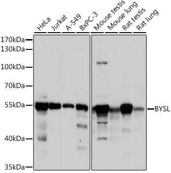 Western blot analysis of extracts of various cell lines, using BYSL antibody at 1:1000 dilution.
Secondary antibody: HRP Goat Anti-Rabbit IgG at 1:10000 dilution.
Lysates/proteins: 25ug per lane.
Blocking buffer: 3% nonfat dry milk in TBST.
Detection: ECL Basic Kit .
Exposure time: 10s. -
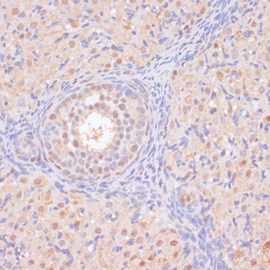 Immunohistochemistry of paraffin-embedded rat ovary using BYSL antibody at dilution of 1:100 .Perform microwave antigen retrieval with 10 mM PBS buffer pH 7.2 before commencing with IHC staining protocol.
 -
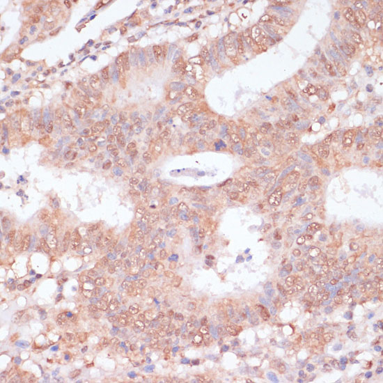 Immunohistochemistry of paraffin-embedded rat ovary using BYSL antibody at dilution of 1:100 .Perform microwave antigen retrieval with 10 mM PBS buffer pH 7.2 before commencing with IHC staining protocol.
 -
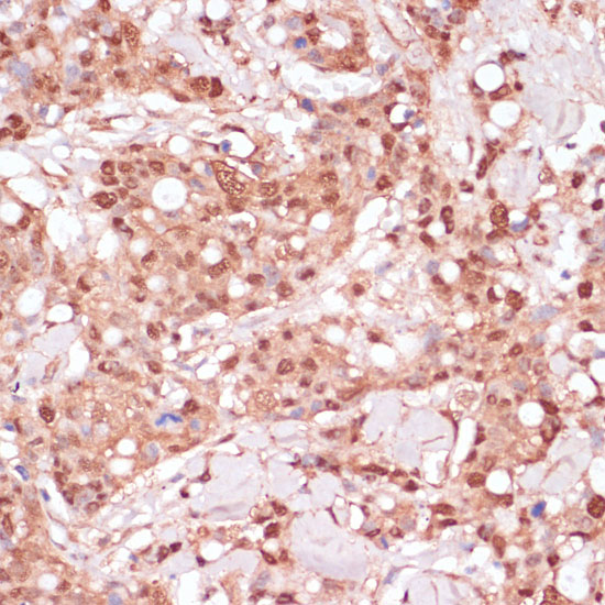 Immunohistochemistry of paraffin-embedded rat ovary using BYSL antibody at dilution of 1:100 .Perform microwave antigen retrieval with 10 mM PBS buffer pH 7.2 before commencing with IHC staining protocol.
Bioworld Biotech only provide peptides for our antibodies and do not provide additional peptide customization services.
Price/Size :
USD 368/1mg/vial

Tips:
For phospho antibody, we provide phospho peptide(0.5mg) and non-phospho peptide(0.5mg).Describe :
Blocking peptides are peptides that bind specifically to the target antibody and block antibody binding. These peptide usually contains the epitope recognized by the antibody. Antibodies bound to the blocking peptide no longer bind to the epitope on the target protein. This mechanism is useful when non-specific binding is an issue, for example, in Western blotting (WB) and Immunohistochemistry (IHC). By comparing the staining from the blocked antibody versus the antibody alone, one can see which staining is specific; Specific binding will be absent from the western blot or IHC performed with the neutralized antibody.Formula:
Synthetic peptide was lyophilized with 100% acetonitrile and is supplied as a powder. Reconstitute with 0.1 ml DI water for a final concentration of 10 mg/ml.The purity is >90%,tested by HPLC and MS.
Storage:
The freeze-dried powder is more stable. For short time at 2-8°C. For long term storage store at -20°C.
Note :
This product is for research use only (RUO only). Not for use in diagnostic or therapeutic procedures.